Elbow Disorders of the
Cavalier King Charles Spaniel
-
 Incomplete
Ossification
Incomplete
Ossification - Elbow Fractures
- Elbow Luxation & Elbow Dysplasia
- Research News
- Related Links
- Veterinary Resources
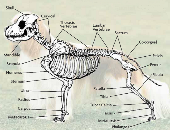
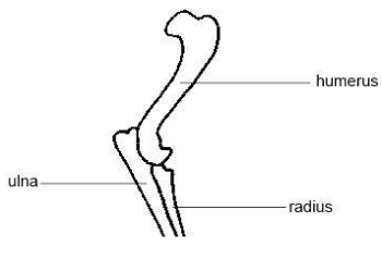
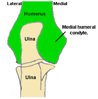
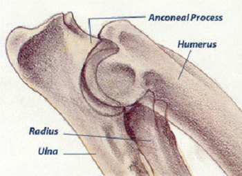
The elbow of the cavalier King Charles spaniel (CKCS) is a delicately
structured combination of moving bones: the
humerus,
which extends from the shoulder, meets and joins the two forearm
bones, the radius and the ulna, at the
elbow joint. The knuckle-shaped tip of the humerus bone is called the
condyle. In fact, the
 word condyle comes from the Greek
word for knuckle.
word condyle comes from the Greek
word for knuckle.
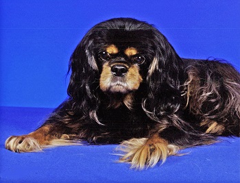
As the photo at the right shows, the condyle has two large rounded ridges -- the medial aspect, which is closest to the dog's torso, and the lateral aspect, which is farthest from the torso. In the photo, the medial aspect is the ridge on the left and the lateral aspect is the ridge on the right. The small part of that knuckle which connects the median and lateral aspects of the condyle is called the trochlea, and the hole above the trochlea and between the median and lateral aspects is called the supratrochlear foramen or just the foramen.
 Courtesy of The Dog Channel
RETURN TO TOP
Incomplete Ossification
In the cavalier and a few other breeds of dogs (particularly other spaniels, especially springers, Brittanys, and cockers, along with French bulldogs, Labrador retrievers, and Yorkshire terriers), the lower middle section of the condyle -- the trochlea -- may not grow to full maturity during the development months of puppyhood. As a result, there remains a very narrow vertical gap -- a fissure line -- in the trochlea, leading to the hole above it, the foramen. This thin gap -- a fibrous band -- between the median and lateral aspects is due to the failure of the growth plates of the condyle to fuse, what is called incomplete ossification. In this case, it's full name is incomplete ossification of the humeral condyle (IOHC). It creates a weak area predisposing for development of condylar fractures.
IOHC may be a heritable condition in cavaliers, due to the apparent
predisposition to fracturing. In the normal
 canine
condyle, the ossification is complete within 12 weeks after birth. IOHC may amount to a life-long weakness in the affected dog's elbow.
While it is not actually a fracture itself (since it never fused to
begin with), IOHC is a serious defect and may lead to a fracture of the
condyle during even mild trauma against it. IOHC's symptoms, diagnosis,
and treatment are discussed below in those subsections under the
Elbow
Fractures section of this webpage. IOHC was first identified by Dr.
Denis Marcellin-Little (right) in a
November 1994 article.
canine
condyle, the ossification is complete within 12 weeks after birth. IOHC may amount to a life-long weakness in the affected dog's elbow.
While it is not actually a fracture itself (since it never fused to
begin with), IOHC is a serious defect and may lead to a fracture of the
condyle during even mild trauma against it. IOHC's symptoms, diagnosis,
and treatment are discussed below in those subsections under the
Elbow
Fractures section of this webpage. IOHC was first identified by Dr.
Denis Marcellin-Little (right) in a
November 1994 article.
RETURN TO TOP
Elbow Fractures
 What It Is
Cavaliers appear to have a breed predisposition for fractures of their elbows, at the base of the humerus bone. These fractures are called humeral condylar fractures.
What It Is
 Most of these condylar fractures in cavaliers are believed to be
stress fractures or fatigue fractures attributed to either (a) IOHC (described
above), or (b) "progressive humeral intracondylar fissure" (HIF)
which is said to develop the separation of the condyles after normal ossification
has occurred. Otherwise, HIF appears to be very similar to IOHC. See
this
October 2011 article.
Most of these condylar fractures in cavaliers are believed to be
stress fractures or fatigue fractures attributed to either (a) IOHC (described
above), or (b) "progressive humeral intracondylar fissure" (HIF)
which is said to develop the separation of the condyles after normal ossification
has occurred. Otherwise, HIF appears to be very similar to IOHC. See
this
October 2011 article.
The condylar fracture occurs mainly in immature dogs -- usually under 12 month of age -- and depending upon which published veterinary study is reviewed, mainly in French bulldogs, spaniels in general, especially springers, Brittanys, and cockers as well as CKCSs, followed by Labrador retrievers and Yorkshire terriers.
Four different types of humeral condylear fractures have been observed:
(1) Incomplete Ossification (IOHC): The least severe type is not really a fracture at all, but is visible on x-ray as a fissure line within the condyle, indicating that the bone has not broken but that the condyle has not completely ossified, leaving a very narrow horizontal gap. See the section above titled "Incomplete Ossification".
(2) Lateral Condylar Fracture: This is the most common type, with only one ridge of the humerus condyle broken, this one called the "lateral" ridge.
(3) Medial Condylar Fracture: This fraction also is of only one ridge, being called the medial ridge.
(4) Intercondylar Fracture: This type of fracture is of both ridges, and are called "Y" or "T" fractures, depending upon the shape of the breaks.
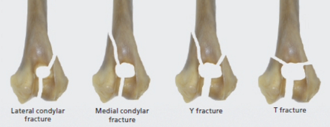 Diagram
courtesy of Dr. Andy Moores, In Practice (2006) 28:391.
Diagram
courtesy of Dr. Andy Moores, In Practice (2006) 28:391.
RETURN TO TOP
Symptoms
Outward signs of elbow fractures are as might be expected, including lameness, indications of pain, a limited range of motion at the elbow (ROM), holding the leg up, and swelling. However, often with these types of fractures, and especially with only incomplete ossification and no real breakage, the dog may not experience any pain at all and show no other typical symptoms except possibly some mild lameness.
Fractures resulting from relatively minor traumas are considered more likely to be associated with IOHC than breaks due to more serious impacts.
RETURN TO TOP
Diagnosis
 Diagnostic
radiology is recommended for any cavalier with otherwise undiagnosed
lameness, even if a physical examination indicates a normal elbow.
Incomplete ossification of the condyle may be a heritable condition in
cavalier King Charles spaniels, due to the apparent predisposition to
fracturing. In the normal canine condyle, the ossification is complete
within 12 weeks after birth. X-rays of the elbow show whether there is
complete ossification or not.
Diagnostic
radiology is recommended for any cavalier with otherwise undiagnosed
lameness, even if a physical examination indicates a normal elbow.
Incomplete ossification of the condyle may be a heritable condition in
cavalier King Charles spaniels, due to the apparent predisposition to
fracturing. In the normal canine condyle, the ossification is complete
within 12 weeks after birth. X-rays of the elbow show whether there is
complete ossification or not.
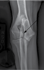
Radiographs (x-rays) are the first line of diagnosis of either IOHC prior to a fracture or the actual fracture itself. X-rays of IOHC before an actual fracture can be hit-or-miss. The fissure line would show up on x-rays as a thin black line, called a radiolucent line extending through the condyle. In the x-ray image at the right here, the black arrow points to a radiolucent line across the humerus condyle, indicating IOHC. [Image is courtesy of Dr. Felix Michael Duerr, Canine Lameness (2020) Fig. 14.4(A)]
If IOHC is suspected in an x-ray prior to a fracture, a means of confirming the diagnosis is to also x-ray the elbow of the unaffected foreleg for comparison purposes. Also, since IOHC may be in both elbows, x-raying both forelegs is always advisable.
The next step in the diagnostic protocol is to use advanced imaging, in the form of either computed tomography (CT) or magnetic resonance imaging (MRI). CT is preferred because it is considered an ideal imaging modality for IOHC and usually is significantly less costly and more readably available than is MRI.
RETURN TO TOP
Treatment
- Medication
- Surgery
- Low-intensity pulsed ultrasound (LIPUS)
- Recombinant bone morphogenetic protein (rhBMP2)
• Medication
IOHC without a fracture may be treated with pain medications if the dog displays signs of being in pain.
Some specialists object to treating IOHC only with medications because of the inherent frailty of the condyle where its growth plates have failed to fuse during maturation.
• Surgery
Surgical insertion of stainless steel or titanium bone screws, compression plates, and Kirschner wire (K-wire) is a standard treatment for IOHC and resulting fractures of the humerus condyle to stabilize the elbow. To minimize complication rates, fractures that are associated with IOHC are typically stabilized using the largest transcondylar screw that can be safely accommodated in the humeral condyle. Post-operative complications and wound infections are not unusual in surgical repairs of elbow breaks due to IOHC. See this February 2015 article and this March 2017 article, and this April 2022 article, and this September 2022 article, for more details about IOHC surgeries.
• Low-intensity pulsed ultrasound (LIPUS)
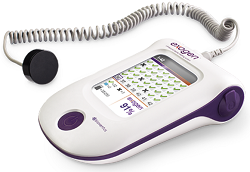 Exogen
is a non-invasive treatment of bone defects (excluding vertebra and the
skull) that includes treatment of delayed unions and non-unions, such as
IOHC, as well as stress fractures and joint fusion. Exogen activates
the biological healing response at the molecular level. Twenty minutes
each day, low-intensity pulsed ultrasound waves from the Exogen unit
(right) pass through the skin and soft tissue to reach the bone at
the point of fracture and accelerate the formation of bone. It
accelerates fracture healing time and repair. Exogen has been proven
effective for healing 86% of nonunion fractures when used as prescribed.
Exogen
is a non-invasive treatment of bone defects (excluding vertebra and the
skull) that includes treatment of delayed unions and non-unions, such as
IOHC, as well as stress fractures and joint fusion. Exogen activates
the biological healing response at the molecular level. Twenty minutes
each day, low-intensity pulsed ultrasound waves from the Exogen unit
(right) pass through the skin and soft tissue to reach the bone at
the point of fracture and accelerate the formation of bone. It
accelerates fracture healing time and repair. Exogen has been proven
effective for healing 86% of nonunion fractures when used as prescribed.
The manufacturer of Exogen represents that there are no contraindications for the Exogen device in humans. However, its safety and effectiveness have not been established for persons lacking skeletal maturity, pregnant or nursing women, patients wit cardiac pacemakers, or fractures due to bone cancer, or on patients with poor blood circulation or clotting problems.
For more information about Exogen, see this website.
• Recombinant bone morphogenetic protein (rhBMP2)
Recombinant bone morphogenetic protein (rhBMP2) is a molecule that enhances ossification.
RETURN TO TOP
Breeders' Responsibilities
Incomplete ossification of the condyle may be a heritable condition in cavalier King Charles spaniels, due to the apparent predisposition to fracturing. In the normal canine condyle, the ossification is complete within 12 weeks after birth. X-rays of the elbow show whether there is complete ossification or not. In a seminal study of this condition, the investigators stated:
"Because the disease may have a genetic basis, we recommend that affected dogs and their sires and dams not be bred. Elbow joints of the littermates of affected dogs that are maintained for breeding should be evaluated radiographically. The coefficient of inbreeding should be kept low in affected families. Because fractures occur mostly in middle-aged dogs, affected dogs may have had offspring before a humeral condyle with incomplete ossification was diagnosed, making control of this disease difficult."
RETURN TO TOP
Elbow Luxation & Elbow Dysplasia
Elbow luxation and elbow dysplasia are two distinct genetic disorders of dogs. While neither of them is particularly common among cavalier King Charles spaniels, a few cases in the breed have been reported.
 Luxation: Congenital elbow luxation is a
polygenic genetic disorder. Cavaliers as a breed are not
known to be predisposed to this disease. All bone elements of the elbow
joint are present in elbow luxation, and relatively normal in size
and shape. However, typically, the radius and the ulna of the elbow
joint are found to be displaced. There are three grades of luxation.
Type I: dislocation of the lateral or caudal aspect of the radial head
Type II: changes in the humeroulnar joint with lateral dislocation of
the proximal ulna. Type III: congenital luxation with displacement of
both the radius and ulna, which is often associated with generalized
joint laxity. Type III is often associated with multiple skeletal
deformities.
Luxation: Congenital elbow luxation is a
polygenic genetic disorder. Cavaliers as a breed are not
known to be predisposed to this disease. All bone elements of the elbow
joint are present in elbow luxation, and relatively normal in size
and shape. However, typically, the radius and the ulna of the elbow
joint are found to be displaced. There are three grades of luxation.
Type I: dislocation of the lateral or caudal aspect of the radial head
Type II: changes in the humeroulnar joint with lateral dislocation of
the proximal ulna. Type III: congenital luxation with displacement of
both the radius and ulna, which is often associated with generalized
joint laxity. Type III is often associated with multiple skeletal
deformities.
In a November 2004 article, a 7-week-old female cavalier was evaluated for non-weight-bearing lameness of the right forelimb. She was diagnosed with Type III unilateral congenital elbow luxation by x-ray. After surgical temporary placement of a transarticular pin, and external splinting of the joint, she was able to bear full weight.
Dysplasia: Elbow dysplasia also is an inherited polygenic disease of the elbow, but inlike luxation, it can involve abnormal developments of the elbow joint (coronoid disease) including fragmentation and/or inflammation (osteochondritis), and/or an un-unitd anconeal process. (See diagram above.) In this February 2017 article, the investigators reported finding "the Cavalier King Charles spaniel had the lowest heritability at 0.01." More details about canine elbow dysplasia are available on the OFA.org website.
RETURN TO TOP
Research News
April 2020:
Cavaliers had 4% of humeral condylar fractures in a three year
UK study.
 In an
April 2020 article, UK veterinary researchers (Matthew A. J. Smith
[right], G.
Jenkins, B. L. Dean, T. M. O'Neill, N. J. Macdonald) reviewed the
medical records at three specialist veterinary centers of 115 dogs under
the age of 12 months examined and treated for humeral condylar fracture
during the period from 2015 to 2018. Of the 115 dogs with 118 humeral
condylar fractures, French bulldogs (41%) and English springer spaniels
(15%) were overrepresented, with cavalier King Charles spaniels
comprising 4%. Lateral condylar fractures occurred in 70% of the cases,
with medial condylar fractures and Y and T fractures accounting for 9%
and 21%, respectively. The median age at the time of fracture was 4
months, and the range was from 2 to 10 months.
In an
April 2020 article, UK veterinary researchers (Matthew A. J. Smith
[right], G.
Jenkins, B. L. Dean, T. M. O'Neill, N. J. Macdonald) reviewed the
medical records at three specialist veterinary centers of 115 dogs under
the age of 12 months examined and treated for humeral condylar fracture
during the period from 2015 to 2018. Of the 115 dogs with 118 humeral
condylar fractures, French bulldogs (41%) and English springer spaniels
(15%) were overrepresented, with cavalier King Charles spaniels
comprising 4%. Lateral condylar fractures occurred in 70% of the cases,
with medial condylar fractures and Y and T fractures accounting for 9%
and 21%, respectively. The median age at the time of fracture was 4
months, and the range was from 2 to 10 months.
April 2020:
Cavaliers had a high prevalence of elbow fractures in an 8-year
UK hospital study.
 In
an
April 2020 article, UK veterinary researchers (Carlos Sanchez
Villamil, Andrew S. J. Phillips, Camilla L. Pegram, Dan G. O'Neill,
Richard L. Meeson [right]) reviewed the medical records of 112
dogs diagnosed with humeral condylar fractures (HCF) -- breaks of the
upper foreleg bone at the elbow -- at the Royal Veterinary College's
Queen Mother Hospital for Animals during the period from January 2010
through August 2018, totaling records of 43,325 dogs overall. The The
breeds with the highest HCF prevalence were French bulldog 2.40%,
English springer spaniel 2.25%, cocker spaniel 0.62%, and cavalier King
Charles spaniel 0.47%.
In
an
April 2020 article, UK veterinary researchers (Carlos Sanchez
Villamil, Andrew S. J. Phillips, Camilla L. Pegram, Dan G. O'Neill,
Richard L. Meeson [right]) reviewed the medical records of 112
dogs diagnosed with humeral condylar fractures (HCF) -- breaks of the
upper foreleg bone at the elbow -- at the Royal Veterinary College's
Queen Mother Hospital for Animals during the period from January 2010
through August 2018, totaling records of 43,325 dogs overall. The The
breeds with the highest HCF prevalence were French bulldog 2.40%,
English springer spaniel 2.25%, cocker spaniel 0.62%, and cavalier King
Charles spaniel 0.47%.
RETURN TO TOP
Related Links
RETURN TO TOP
Veterinary Resources
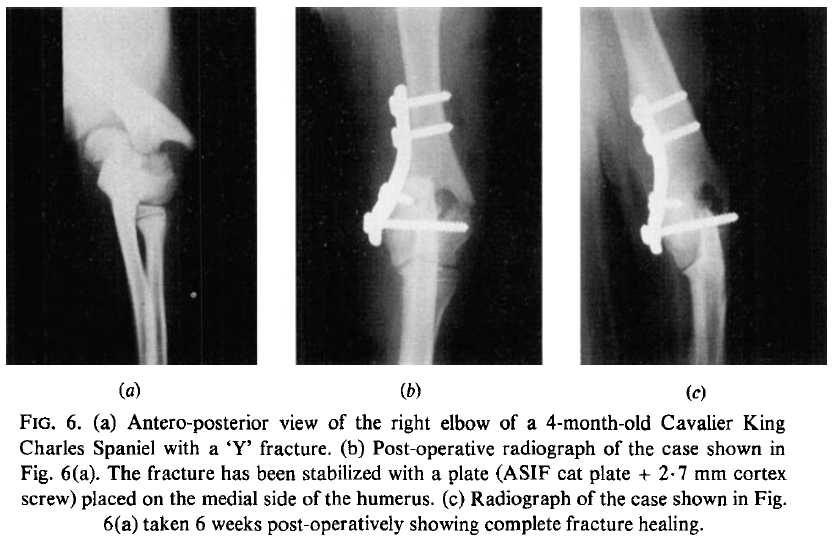
Condylar fractures of the humerus in the dog; a review of 133 cases. H. R. Denny. J. Small Anim. Pract. 1983;24:185-197. Quote: Case histories have been reviewed of 133 dogs with condylar fractures of the humerus. The fractures were divided into three types: lateral condylar, medial condylar and intercondylar. For each type the breed and age prevalence, the cause of fracture, the method of fixation and the results of treatment have been recorded. ... Seventy-four dogs had fractures of the lateral condyle. The injury was seen in 20 breeds and Spaniels (38 per cent) were most frequently affected [including 5 Cavalier King Charles Spaniels]. The sex ratio was equal. Sixty-seven per cent of cases were under a year of age and the peak age prevalence was 4 months (Table 2). The commonest cause of fracture was a fall; other causes are listed in Table 3. In 65 dogs the fracture was stabilized with a single lag screw driven through the lateral condyle into the medial condyle." See Figure 6 (below) of the right elbow of a 4-month-old cavalier.
Incomplete Ossification of the Humeral Condyle in Spaniels. Denis J. Marcellin-Little, David J. Deyoung, Kelli K. Ferris, Clifford M. Berry. Vet. Surg. November 1994; 23(6):475-487. Quote: An evaluation of 157 dogs with humeral fractures was performed. Cocker spaniels were more likely to have humeral condylar fractures (HCFs) than other breeds. Male cocker spaniels were at increased risk. Cocker spaniels had more bilateral HCFs than other breeds of dogs. Eighteen dogs (17 purebred spaniels and 1 crossbred spaniel) with HCFs of unknown cause or occurring with normal activity were further studied, using radiography of their humeral condyle bilaterally (n = 18), computed tomography (n = 3), biopsy (n = 2), bone scintigraphy (n = 2), and genetic evaluation (n = 8). Fourteen of these 18 dogs had a nonfractured contralateral condyle. Twelve (86%) of the 14 nonfractured humeral condyles had a radiolucent line within the center of the condyle, 13 (93%) had radiographic signs of degenerative joint disease and an abnormal medial coronoid process, and six (43%) had periosteal proliferation involving the lateral epicondyle. Examination of biopsy samples from the fracture sites of two cocker spaniels showed fibrous tissue present at the fracture surfaces. The results of this study suggest an association between incomplete ossification of the humeral condyle in cocker spaniels and Brittany spaniels and a high prevalence of HCFs. Eight affected cocker spaniels with available pedigree information were found to be genetically related, suggesting that incomplete ossification of the humeral condyle may be a genetic disease with a recessive mode of inheritance.
Triceps Tenotomy and Double Plate Stabilization of "Y-T" Fracture of the Humeral Condyle in Three Dogs. C. Sturgeon, A. M. Wilson, P. McGuigan, T. J. Lawes, P. Muir. Vet. Comp. Orthop. Traumatol. 2000;13:34-38. Quote: Triceps tenotomy was used for open reduction of "Y-T" fractures of the humeral condyle in three dogs [including 1 cavalier King Charles spaniel]. Stabilization of the fracture with a bone screw inserted for lag effect and use of a bone plate on each epicondylar crest resulted in satisfactory healing of the fracture and good limb function in all of the patients.
Incomplete ossification of the humeral condyle in two Labrador retrievers. D. Robin, D. J. Marcellin-Little. J. Sm. Anim. Pract. May 2001;42:231-234. Quote: Incomplete ossification of the humeral condyle (IOHC) has been reported in specific breeds: cocker spaniels, Brittany spaniels, springer spaniels, Cavalier King Charles spaniels and clumber spaniels, as well as in a pug and a rottweiler. IOHC was identified in two Labrador retrievers using computed tomography. Both dogs were non-weight bearing on the affected forelimbs. The dogs were treated by means of a bone screw placed across the humeral condyle. IOHC was originally reported in spaniel and chondrodystrophic breeds. The pathogenesis of the condition remains unknown, but may be related to impaired antebrachlal bone growth, similarly to the pathogeneses of elbow dysplasia and radius curvus.
Unilateral congenital elbow luxation in a Cavalier King Charles Spaniel. Heather L. McDonell. Can Vet J. November 2004;45(11):941-943. Quote: "A 7-week-old, intact female, Cavalier King Charles Spaniel was evaluated for nonweight bearing lameness of the right forelimb. Type III unilateral congenital elbow luxation was diagnosed radiographically. After surgical reduction, temporary placement of a transarticular pin, and external splinting of the joint, full weight bearing was achieved. Radial head subluxation persisted."
Bilateral fixation of Y-T humeral condyle fractures via medial and lateral approaches in 29 dogs. W. M. McKee, C. Macias, J. F. Innes. J. Sm. Anim. Pract. May 2005;46:217-226. Quote: OBJECTIVES: To describe bilateral fixation of Y-T fractures of the humeral condyle via combined medial and lateral approaches, and to determine the technique's clinical and radiographic short-term outcomes. METHODS: Details of 30 consecutive fractures in 29 dogs were reviewed [including 4 cavalier King Charles spaniels]. These included signalment, method of fixation, complications, and follow-up limb function and range of elbow joint motion. RESULTS: The age of the dogs ranged from three months to nine years, and bodyweight ranged from 1·9 to 48 kg. The humeral condyle was reattached to the shaft using medial and lateral bone plates in 18 fractures, a medial plate and lateral Kirschner wire(s) in six fractures, and medial and lateral Kirschner wire(s) in six fractures. Major complications were recorded in four fractures and minor complications in two fractures. Limb function at follow-up was graded as excellent in 12, good in 15 and fair in three fractures. The range of elbow flexion was normal in seven, mildly reduced in 18, moderately reduced in four and severely reduced in one fracture. CLINICAL SIGNIFICANCE: In contrast to the caudal approach, combined medial and lateral approaches decrease the extent of periarticular soft tissue dissection, avoid complications associated with olecranon osteotomy and enable exposure of the entire humeral diaphysis for fixation. Bilateral fixation is likely to be better at counteracting bending and torsional forces compared with unilateral fixation.
Evaluation of elbow incongruency using reconstructed CT in dogs suffering fragmented coronoid process. T. J. Gemmill, D. J. Mellor, D. N. Clements, S. P. Clarke, M. Farrell, D. Bennett, S. Carmichael. J. Sm. Anim. Pract. July 2005;46:327-333. Quote: OBJECTIVES: A retrospective study was undertaken to evaluate elbow joint congruency in dogs suffering fragmented coronoid process (FCP). METHODS: Based on clinical, radiographic and computed tomographic (CT) examinations, elbows were divided into control and FCP groups. Standardised CT reconstructions were formatted in the frontal and sagittal planes. Humeroradial and humeroulnar joint space measurements were obtained from the images and incongruencies were calculated by comparing the two measurements. RESULTS: Forty-two FCP and 29 control elbows [including 3 cavalier King Charles spaniels] were identified. No incongruencies were noted at the coronoid base. At the level of the coronoid apex, FCP elbows exhibited a significant radioulnar incongruency compared with controls, though incongruency was not identified in all cases. Comparing FCP and control elbows at the level of the apex, the humeroradial joint space was increased in FCP elbows (P=0·0006) whereas no difference was noted in the humeroulnar space. CLINICAL SIGNIFICANCE: This study supports the hypothesis that joint incongruency is associated with FCP in dogs, though is not present in every case at the time of diagnosis. The precise mechanism of development of this incongruency cannot be determined from these data.
Treatment of Incomplete Ossification of the Humeral Condyle with Autogenous Bone Grafting Techniques. Noel Fitzpatrick, Thomas J. Smith, Jerry O'Riordan, Russell Yeadon. Vet. Surg. 2009; doi: 10.1111/j.1532-950X.2008.00485.x. Quote: Objective--To report clinical experience with autogenous bone grafting, with and without metallic implants, for treatment of lameness attributed to incomplete ossification of the humeral condyle (IOHC). Study Design--Case series. Animals--Dogs (n=8; 9 elbows [including 2 cavalier King Charles spaniels]) with IOHC. Methods--A transcondylar humeral bone core was removed and the resultant socket grafted using autogenous bone harvested as either free cancellous bone or a corticocancellous dowel using an osteochondral autograph transfer system. In 8 elbows, additional support for the humeral condyle was provided with metallic implants. Postoperative outcome was assessed by clinical, radiographic, computed tomographic (CT) and owner questionnaire examinations in the short and medium term. Results--Eight dogs (9 elbows [including 2 CKCSs]) were treated surgically for IOHC. Graft types were free cancellous graft (n=2) or corticocancellous dowel (7). Condylar augmentation was performed using epicondylar cross pins (1); transcondylar AcutrakTM (AT) screw and epicondylar cortical screw (1); and a single transcondylar AT screw (7). Lameness resolved in 1-12 weeks. Bone bridging was documented in 7 of 8 elbows assessed by CT examination. Owner questionnaires (6 dogs) assessing daily functions were available for 7 of 9 elbows (follow-up, 6-45 months). Relevant follow-up function scores were significantly improved compared with preoperative values. One dog was intermittently lame and was administered nonsteroidal antiinflammatory medication. Conclusion--Use of autogenous bone grafting techniques leads to resolution of lameness attributed to IOHC. Augmentation of grafts with implants like the AT screw is recommended. Clinical Relevance--Autogenous bone grafting techniques represent a viable alternative or adjunct to existing techniques for clinical management of IOHC in the dog.
Postoperative Complications after Surgical Management of Incomplete Ossification of the Humeral Condyle in Dogs. Rachel Hattersley, Malcolm McKee, Turlough O'Neill, Stephen Clarke, Steven Butterworth, Thomas Maddox, Martin Owen, Sorrel J. Langley-Hobbs, Eithne Comerford. Vet. Surg, 2011; doi: 10.1111/j.1532-950X.2011.00847.x. Quote: Objective: To describe incidence and type of postoperative complications in the surgical management of incomplete ossification of the humeral condyle (IOHC) and identify any risk factors associated with development of these complications. Study Design: Case series. Methods: Clinical records of dogs (n=57 [including cavalier King Charles spaniels]) that had prophylactic transcondylar screw insertion for treatment of IOHC (79 elbows) at 6 UK referral centers were reviewed. Signalment, presentation, surgical management, postoperative care, and complications were recorded. Postoperative complications were divided into seroma, surgical site infections (SSI) and implant complications. Results: Spaniel breeds and entire males were overrepresented. The overall complication rate was 59.5%. Seroma (n=25) and SSI (24) were the most commonly encountered complications. Implant failure occurred in 2 dogs. Labrador retrievers were at greater risk of developing a postoperative complication than other breeds (P=.03). Increasing bodyweight was a significant risk factor for development of a SSI (P=.03). Placement of the transcondylar screw in lag fashion rather than as a positional screw reduced the incidence of postoperative SSI (P=.007). Conclusions: Surgical management of IOHC is associated with a high rate of postoperative complications. Placement of the transcondylar screw in lag fashion may limit postoperative complications and warrants further consideration.
Computed Tomographic Documentation of the Natural Progression of Humeral Intracondylar Fissure in a Cocker Spaniel. Michael Farrell, Tim Trevail, William Marshall, Russell Yeadon, Stuart Carmichael. Vet. Surg. October 2011;40:966-971. Quote: Objective: To report the computed tomographic (CT) documentation of humeral intracondylar fissure (HIF ) developing after complete ossification of the humeral condyle (HC). Study Design: Clinical report. Animals: Male 3 year old working (English) Cocker Spaniel. Methods: Sequential CT screening (659‐day interval between analyses). Results: A sagittal hypodense fissure typical of incomplete ossification of the humeral condyle (IOHC ) was identified ∼22 months after screening CT examination documented a normal elbow joint. Conclusion: Even in dogs with clinical features typical of the condition most commonly termed IOHC , fissure formation and propagation can occur after ossification is complete.
Magnetic Resonance Imaging Features of Canine Incomplete Humeral Condyle Ossification. Valentina Piola, Barbara Posch, Heidi Radke, Gerard Telintelo, Michael E. Herrtage. Vet. Radio. & Ultrasound. March 2012; doi: 10.1111/j.1740-8261.2012.01941.x. Quote: Incomplete ossification of the humeral condyle (IOHC) is characterized by an intracondylar fissure located where the intercondylar physis is present in growing dogs. Its radiologic and computed tomographic features have been described but the magnetic resonance (MR) features have not been characterized. Our purpose was to further describe the range o fMR appearances of IOHC, to assess the diagnostic capability ofMR relative to radiology, and to determine whether MR is able to identify the disease before a fissure forms. Thirty-eight elbow MR scans and radiographs, when available, were reviewed and divided into three groups. In Group 1 (affected elbows, n = 22 [including 1 cavalier King Charles spaniel]), there was an intracondylar defect on MR with variable appearance; the defect was not visible radiographically in 32% of the elbows. The main difference between Group 2 (nonaffected elbows, n = 6) and Group 3 (contralaterals to IOHC or to condylar fracture, without fissure, n = 10 [including 1 CKCS]) was the appearance of the humeral condyle in short tau inversion recovery (STIR) sequences: all elbows in Group 2 had a homogeneous humeral condyle, whereas all but one in Group 3 were heterogeneous.One dog in Group 3 developed a complete condylar fissure 7 months after the first examination, when no evidence of an intracondylar defect had been detected. The MR appearance of IOHC is variable and a heterogeneous humeral condyle in STIR images without a clear defect may warn of the possibility for the subsequent development of a condylar fissure.
Effect of Fixation Method on Postoperative Complication Rates After Surgical Stabilization of Lateral Humeral Condylar Fractures in Dogs. Karen L. Perry, Mieghan Bruce, Samantha Woods, Clare Davies, Laura A. Heaps, Gareth I. Arthurs. Vet, Surg. February 2015; doi: 10.1111/j.1532-950X.2014.12276.x. Quote: Objectives: To assess the impact of stabilization method on the complication rate after lateral humeral condylar fracture (LHCF) repair. Study Design: Retrospective multicenter clinical cohort study. Animals: Dogs (n=151) with LHCF [including 7 cavalier King Charles spaniels]. Methods: Medical records (2004-2012) were reviewed for dogs that had surgical repair of LHCF. Data retrieved included signalment, cause of fracture, evidence of incomplete ossification of the humeral condyle, occurrence of postoperative complications, presence of supracondylar comminution preoperatively, and persistence of an intracondylar fissure postoperatively. Outcome was assessed based on the most recent data available and graded as excellent, good, fair, or poor. Results: LHCF (n¼135) were evaluated in 132 dogs; 61 fractures were stabilized using a transcondylar screw and supracondylar K‐wire, 13 using a transcondylar screw and supracondylar screw, and 61 using a transcondylar screw and lateral epicondylar plate. Major complications were significantly (P¼.01) more common after stabilization using a transcondylar screw and supracondylar K‐wire (28%) than in dogs where a supracondylar screw or lateral epicondylar plate were used (11%). Cases that had postoperative complications were significantly (P¼.02) more likely to have a poor outcome. Conclusions: LHCF stabilized using a transcondylar screw and supracondylar K‐wire are more likely to have major complications resulting in a poorer outcome than cases stabilized using a supracondylar screw or lateral epicondylar plate.
Intraosseous stress distribution and bone interaction during load application across the canine elbow joint: A preliminary finite element analysis for determination of condylar fracture pathogenesis in immature and mature dogs. Beatrice Bohme, Vinciane d'Otreppe, Jean-Phillippe Ponthot, Marc Balligan. Research in Vet. Sci. June 2016;106:143-148. Quote: "Distal humeral fractures are common fractures especially in immature small breed dogs. The pathogenesis is still unknown. ... A breed predisposition [of condylar fractures] seems to be present in Yorkshire Terriers, French and English Bulldogs, Pinscher, Springer Spaniels, Cocker Spaniels and Cavalier King Charles Spaniels... For this study, a three- dimensional bone model of the canine elbow was created and finite element analysis performed in order to determine the relationship between fracture type and bone interactions. Fused and non-fused humeral condyles were considered. A failure criterion was implemented to simulate the pathogenesis until fracture. Our study results confirm the clinical observation that lateral condylar fracture is the most common fracture type, implying interaction with the radius. Medial and Y-fractures are less common and occur always in interaction with the ulna whereas the radius causes lateral condylar fracture. Additionally, the fracture type is sensitive to bone positioning during trauma. The pathogenesis of distal humeral fractures is more complex than generally reported in the literature. Highlights: • Distal humeral fracture type may be sensitive to bone positioning during trauma. • The pathogenesis of condylar fractures is more complex than expected from literature. • Medial and Y condylar fractures occur after humeroulnar interaction. • Humero-radial interaction causes lateral condylar fractures."
Fragmentation of the Medial Coronoid Process in Toy and Small Breed Dogs: 13 Elbows (2000-2012). Eric C. Hans, W. Brian Saunders, Brian S. Beale, Don A. Hulse. J. Amer. Anim. Hosp. Assn. June 2016. Quote: Fragmentation of the medial coronoid process (FCP) is an uncommon cause of thoracic limb lameness in toy and small breed dogs. Arthroscopic findings and treatment remains poorly described. The objective of this study was to describe the arthroscopic findings and short-term outcome following arthroscopic treatment in toy and small breed dogs with FCP. Medical records were retrospectively reviewed. Arthroscopic findings were available from 13 elbows (12 dogs). Outcome data >4 wk postoperatively were available for nine elbows. Owner satisfaction scores were available for 10 elbows. Common preoperative findings included lameness, elbow pain, and imaging abnormalities consistent with FCP. Displaced FCP was the most common FCP lesion identified. Cartilage lesions at the medial coronoid process were identified in 92.3% of elbows (n = 12), with a median Outerbridge score of 4 (range 1-5). Concurrent cartilage lesions of the medial humeral condyle were identified in 76.9% of elbows (n = 10). Seven of nine elbows had full or acceptable function post-operatively. Median owner outcome satisfaction was 91% (range 10-100). FCP should be considered a cause of thoracic limb lameness in toy and small breed dogs. Arthroscopy can be safely and effectively used to diagnose and treat FCP in these breeds.
Long-term genetic selection reduced prevalence of hip and elbow dysplasia in 60 dog breeds. A. M. Oberbauer, G. G. Keller, T. R. Famula. Plos ONE. February 2017; doi: 10.1371/journal.pone.0172918. Quote: Canine hip dysplasia (CHD) and elbow dysplasia (ED) impact the health and welfare of all dogs. The first formally organized assessment scheme to improve canine health centered on reducing the prevalence of these orthopedic disorders. Phenotypic screening of joint conformation remains the currently available strategy for breeders to make selection decisions. The present study evaluated the efficacy of employing phenotypic selection on breed improvement of hips and elbows using the Orthopedic Foundation for Animals complete database spanning the 1970-2015 time period. Sixty breeds having more than 1000 unique hip evaluations and 500 elbow evaluations (1,056,852 and 275,129 hip and elbow records, respectively) were interrogated to derive phenotypic improvement, sex and age at time of assessment effects, correlation between the two joints, heritability estimates, estimated breeding values (EBV), and effectiveness of maternal/paternal selection. The data demonstrated that there has been overall improvement in hip and elbow conformation with a reduction in EBV for disease liability, although the breeds differed in the magnitude of the response to selection. Heritabilities also differed substantially across the breeds as did the correlation of the joints; in the absence of a universal association of these differences with breed size, popularity, or participation in screening, it appears that the breeds themselves vary in genetic control. ... The Welsh springer spaniel had an estimated ED heritability of 0.90 ± 0.04 whereas the Cavalier King Charles spaniel had the lowest heritability at 0.01 ± 0.01. ... The five breeds considered to be at low risk for ED are Boxer, Flat coated retriever, Bichon, Cavalier King Charles spaniel and Briard. ... There was subtle, though again breed specific, impact of sex and older ages on CHD and ED. There was greater paternal impact on a reduction of CHD. In the absence of direct genetic tests for either of these two diseases, phenotypic selection has proven to be effective. Furthermore, the data underscore that selection schemes must be breed specific and that it is likely the genetic profiles will be unique across the breeds for these two conditions. Despite the advances achieved with phenotypic selection, incorporation of EBVs into selection schemes should accelerate advances in hip and elbow improvement.
The natural history of humeral intracondylar fissure: an observational study of 30 dogs. A. P. Moores, A. L. Moores. J. Sm. Anim. Pract. March 2017. Quote: Objectives: To determine the risk of condylar fracture, or of needing to have a transcondylar screw placed, and to identify risk factors in a cohort of dogs with humeral intracondylar fissure (also known as incomplete ossification of the humeral condyle) that was initially managed non-surgically. Methods: A retrospective owner survey of dogs diagnosed with humeral intracondylar fissure as an incidental finding and managed non-surgically with a minimum of two years follow-up. Body weight, age, estimated fissure size, gender and contralateral fracture at the time of diagnosis were evaluated as potential risk factors for the development of a humeral condylar fracture or for having a transcondylar screw placed. Results: Data were available for 30 dogs (34 elbows). ... There were 21 English springer spaniels, five cocker spaniels and one each of cavalier King Charles spaniel, Clumber spaniel, Welsh springer spaniel and Labrador. ... Six humeral condyles with a mean fissure size of 50% fractured at a mean of 14 months after diagnosis. A transcondylar screw was placed across two humeral condyles with fissure sizes of 60 and 100% at 11 and 17 months. No risk factors were identified for fracture/screw placement. For those cases that did not fracture or have a screw placed mean fissure size was 52% and mean follow-up time was 56 months (range 29 to 79 months). Clinical Significance: Eighteen percent of cases progressed to fracture and 24% in total required surgery. This information allows clinicians and owners to make an informed decision regarding surgery when faced with a dog with humeral intracondylar fissure identified as an incidental finding.
Spontaneous Septic Arthritis of Canine Elbows: Twenty-One Cases. Ben Mielke, Eithne Comerford, Kate English, Richard Meeson. Vet Comp Orthop Traumatol. November 2018;31(6):488-493. Quote: Objective: This study provides information on clinical features, diagnosis, treatment and associated risk factors of spontaneous septic elbow arthritis in the dog. Methods: Medical records between March 2007 and June 2015 were searched for cases of spontaneous septic elbow arthritis with a diagnosis based on clinical signs, arthrocentesis, cytological and microbiological analysis of elbow joint synovial fluid, radiography and outcome following treatment. Results: Twenty-one cases of septic arthritis were identified [including one cavalier King Charles spaniel]. Pre-existing osteoarthritis was present in 14/15 elbows for which diagnostic imaging was available. Although all cases had increased neutrophil count on synovial fluid cytology, culture was only positive in 12/21. Despite initial improvement in lameness scores (pre-treatment 9/10 [range: 1-10] versus post-treatment 3/10 [range: 1-5]), 11/12 had residual long-term lameness. Recurrence of infection was noted in 3/12 elbows for which long-term (>8 weeks) follow-up was available. There was an acute mortality rate of 2/21 associated with severe systemic sepsis. Clinical significance: Septic arthritis, even in the absence of pyrexia, should be considered as a major differential diagnosis in middle aged, large breed dogs, with pre-existing elbow arthritis, that suffer an acute onset lameness, with elbow joint effusion and discomfort. Antibiotic therapy alone was effective for treatment with high initial response rates. Chronic lameness post-treatment was common, and a high rate of recurrence was seen with 3/12 dogs suffering more than one episode.
Elbow Region. Felix Michael Duerr. Chapter 14 in Canine Lameness. First ed. Edited by Felix Michael Duerr. John Wiley & Sons. 2020;195-221. Quote: This chapter discusses conditions affecting the elbow region (i.e. any of the structures affecting the elbow joint and surrounding area). A brief discussion of the clinically relevant local anatomy is followed by a detailed description of the history, physical examination, and diagnostic findings typically found in patients presenting with pathology of this region. ... Type II luxations occur more often in small-breed dogs (Rahal et al. 2000). Of the three variants of CEL, Type III luxation occurs less frequently and is poorly described, but it is known to occur in small-breed dogs with a case study in Cavalier King Charles Spaniel reported (McDonell 2004).
Epidemiology and clinical management of elbow joint disease in dogs under primary veterinary care in the UK. Dan G. O'Neill, Dave C. Brodbelt, Rebecca Hodge, David B. Church, Richard L. Meeson. Canine Med. & Genetics. February 2020;doi: 10.1186/s40575-020-0080-5. Quote: Background: Conditions affecting the elbow joint are a common cause of lameness in dogs. Primary-care veterinary clinical data are now recognised as a valuable research resource. Using data from the VetCompass Programme, this study aimed to report the frequency and risk factors for elbow joint disease in dogs under primary veterinary care in the UK and describe clinical management. Results: From 455,069 dogs under veterinary care, the one-year period prevalence for elbow joint disease diagnosis was 0.56% (95% CI: 0.53-0.60). Of 616 incident cases, the most common specific variants of elbow joint disease were osteoarthritis (n = 468, 75.97%), elbow dysplasia (190, 30.84%) and traumatic (41, 6.66%). The most common signs described by the owners were lameness (n = 466, 75.65%), difficulty exercising (123, 19.97%) and pain (86, 13.96%). The most common findings recorded on veterinary examination were pain (n = 283, 45.94%), lameness (278, 45.13%) and reduced range of movement (243, 39.45%). Common medications used included non-steroidal anti-inflammatory drugs (n = 544, 88.31%), tramadol (121, 19.64%) and disease modifying agents (118, 19.16%). Of 109 deaths involving euthanasia with information available from the 616 incident cases, elbow joint disease contributed to the decision to euthanase in 45 (41.28%) dogs. Five breeds showed increased odds of elbow joint disease compared with crossbred dogs: Rottweiler (OR: 6.16, 95% CI 3.89-9.75), Labrador Retriever (OR: 5.94, 95% CI 4.65-7.60), German Shepherd Dog (OR: 4.13, 95% CI 2.88-5.93), Golden Retriever (OR: 3.11, 95% CI 1.93-5.00) and English Springer Spaniel (OR: 2.00, 95% CI 1.26-3.18). ... The current study identified Boxers with an odds ratio of 0.68, although Cavalier King Charles spaniels had an odds ratio of 1.15. ... Additional risk factors included having an adult bodyweight that was equal or higher than their breed/sex mean, advancing age, being male, being neutered, being insured and larger bodyweight. Conclusions: Elbow joint disease is a relatively common diagnosis in dogs and has a high welfare impact as evidenced by the high proportion of cases recorded with pain, lameness and analgesic therapy. There are strong breed predispositions, in particular for large breed dogs. These findings present a clear case for improved breeding programmes to reduce the burden of elbow joint disease.
Impact of breed on canine humeral condylar fracture configuration, surgical management, and outcome. Carlos Sanchez Villamil, Andrew S. J. Phillips, Camilla L. Pegram, Dan G. O'Neill, Richard L. Meeson. Vet. Surg. April 2020; doi: 10.1111/vsu.13432. Quote: Objective: To report the prevalence, configuration, risk factors, fixation methods and outcomes after repair of humeral condylar fractures (HCF) in dogs. Study design: Retrospective nested cohort study. Methods: Medical records of dogs referred between January 2010 and August 2018 were searched for HCF. Demographics, fracture configuration, repair, and complications were obtained from medical records. Radiographs were assessed for fracture reduction, implant positioning, and bone healing. Short-term radiographic and clinical outcomes, and long-term owner-assessed outcome was determined. Associations between these variables were statistically analyzed. Results: Dogs with HCF represented 112 of 43,325 (0.26%) referrals [including 6 cavalier King Charles spaniels (CKCS) - 5.4%]. ... The breeds with the highest HCF prevalence were French bulldog 2.40%, English springer spaniel 2.25%, cocker spaniel 0.62%, Cavalier King Charles spaniel 0.47%, and crossbreeds 0.18%. ... English springer spaniels, French bulldogs, cocker spaniels, and Cavalier King Charles spaniels were predisposed to HCF on multivariable analysis. ... French bulldogs were 6.58 times more likely than other breeds to have a medial HCF. Epicondylar plate fixation was associated with reduced complications com-ared with lag screws and Kirschner wires. Lameness was scored as 1 of 5 (median) in the 85 dogs with initial follow-up (median 6 weeks) after HCF repair. Outcome was considered excellent in 26 of 31 dogs with long-term follow-up (median 36 months). Conclusion: French bulldogs and spaniels were predisposed to HCF, and medial HCF were more common in French bulldogs [and CKCSs]. Epicondylar plate fixation was associated with reduced complications. Clinical significance: French bulldogs are predisposed to HCF, including medial HCF. Epicondylar plate fixation is recommended over other epicondylar fixation methods to reduce complications.
Effect of breed as a risk factor for humeral condylar fracture in skeletally immature dogs. M. A. J. Smith, G. Jenkins, B. L. Dean, T. M. O'Neill, N. J. Macdonald. J. Sm. Anim. Pract. April 2020; doi: 10.1111/jsap.13144. Quote: Objectives: To investigate the effect of breed as a risk factor associated with humeral condylar fracture in skeletally immature dogs in the UK. Materials and Methods: Retrospective study of dogs under 12 months of age that were presented with humeral condylar fracture to three specialist referral centres between 2015 and 2018. Data retrieved from medical records included breed, age, gender, neuter status, affected limb, fracture configuration and aetiology of the fracture. Breed population percentages were compared with those recorded by the UK Kennel Club. Results: Of the 115 dogs with 118 fractures, French bulldogs (41%) and English springer spaniels (15%) were overrepresented [cavalier King Charles spaniels 4%]: humeral condylar fractures were more commonly diagnosed in French bulldogs and English springer spaniels compared with mixed‐breed dogs. Lateral condylar fractures occurred in 70% of cases, with medial condylar fractures and Y/T fractures accounting for 9% and 21%, respectively. Median age at the time of fracture was 4 months (range 2 to 10 months). Clinical Significance: French bulldogs and English springer spaniels were identified as being at potentially increased risk of humeral condylar fracture in skeletally immature dogs.
Bilateral locking compression plate and transcondylar screw fixation for stabilization of canine bicondylar humeral fractures. Javier García, Russell Yeadon, Miguel A. Solano. Vet. Surg. April 2020; doi: 10.1111/vsu.13429. Quote: Objective: To report the surgical management, complications, and short-term outcomes for dogs with humeral Y-T fractures (ie, fractures affecting the medial and lateral aspect of the humeral condyle extending from the articular surface) that had been approached bilaterally (medial and lateral) and stabilized with two locking compression plates (LCP) and to investigate risk factors for major complications in the short term. Study design: Retrospective case series. Animals: Twenty-eight client-owned dogs [including one cavalier King Charles spaniel; 7 French bulldogs, 11 spaniels]. Methods: Medical records of 28 dogs that presented with 29 consecutive Y-T fractures that had been approached bilaterally, reduced, and stabilized by using a transcondylar screw (in lag or positional fashion) and two or three LCP were reviewed. All dogs had at least 6 weeks clinical follow-up, and in some cases, responses from the long‐term owner-based University of Liverpool questionnaire for owners of dogs with mobility problems (Liverpool Osteoarthritis in Dogs) was obtained. Results: Twenty-eight dogs (29 fractures) presented with Y-T fractures and met the inclusion criteria. The fractures were reduced and stabilized with bilateral LCP and screws. The French bulldog (n = 7) was the most commonly represented breed. Age ranged from 4 months to 9.5 years (mean, 43.53±34.63 months) [CKCS was 8 months]; seven (25%) dogs were considered skeletally immature. Twenty-seven (96.4%) dogs were considered to have had resolution of lameness at 6 weeks. Major complications were registered in two (7%) dogs; one dog required surgical revision, and one dog had catastrophic complications leading to amputation-of the limb. The owner-based questionnaire was available for 18 (64%) dogs. Conclusion: Management of canine Y-T humeral fractures stabilized with bilateral LCP and transcondylar screws led to a low complication rate with a good to excellent long‐term outcome according to responses on the client questionnaire. Risk factors for major complications were not identified in our sample population. Clinical significance: Management of Y-T humeral fractures stabilized with bilateral LCP and transcondylar screws can lead to good to excellent short-term outcome with a low complication rate in mature and immature dogs.
Medial epicondylar fissure fracture as a complication of transcondylar screw placement for the treatment of humeral intracondylar fissure. Georgia Jenkins, Andy P. Moores. Vet. Surg. April 2022; doi: 10.1111/vsu.13800. Quote: Objective: To report the incidence of medial epicondylar fissure fracture (MEFF) after medial-to-lateral transcondylar screw placement in dogs with humeral intracondylar fissure (HIF) and to identify risk factors for MEFF. Study design: Retrospective study. Sample population: Seventy-four client-owned dogs (88 elbows). Methods: Medical records of dogs surgically treated for HIF, and postoperative imaging studies were reviewed for demographics, fracture characteristics, and repair techniques. The width of the transcondylar screw was expressed relative to the height of the condyle. Screw angle and degree of countersinking were recorded. Information from case records and follow-up radiographs were used to identify complications. Results: Medial epicondylar fissure fracture was identified in 10 elbows (11.4%) following medial-to-lateral transcondylar screw placement: 4 cases were detected intraoperatively, 2 on immediate postoperative radiographs, 1 during routine radiographic follow up, and 3 when radiographs were reviewed for this study. A larger relative screw size was found to increase the risk of MEFF. Fifteen additional complications were identified in 13/80 elbows at a median of 6 weeks postoperatively (range 1-56 weeks). Screw loosening was the most frequent complication (n = 9) and was the only complication in dogs with MEFF (n = 3); MEFF tended to increase the risk of perioperative screw loosening. Conclusion: Medial epicondylar fissure fracture occurred in 10/88 elbows treated for HIF and was more common in elbows treated with a larger screw size relative to the height of the condyle. Clinical significance: Placing transcondylar screws with a diameter inferior to 41% of the height of the condyle is recommended to avoid MEFF. Medial epicondylar fissure fracture appears to have a low clinical significance in the perioperative period, although its effect on long-term outcome remains unknown.
Humeral unicondylar fractures in immature dogs treated using a bone screw and Kirschner wire. Steven J. Butterworth. Vet. Rec. September 2022; doi: 10.1002/vetr.2176. Quote: Background: To investigate the incidence of implant-related complications when humeral unicondylar fractures in immature dogs are stabilised with a transcondylar bone screw and epicondylar Kirschner wire (K-wire)/pin. Methods: Records of cases treated by the author in this clinic over a 10-year period were screened for those involving humeral condylar fractures. These were then reduced to include those patients who were less than 7 months of age with unicondylar fractures and follow-up radiographs. Results: Thirty fractures in 29 dogs met the inclusion criteria. All of these had been stabilised with a transcondylar bone screw and epicondylar K-wire. Fracture healing was documented in all dogs. Surgical site infection was not recorded. Implant-related major complications were reported in four (13%) dogs and successfully treated by either implant removal or replacement. Short-term outcome was judged as excellent in 23 out of 30 and good in seven out of 30 of these fractures. Conclusion: The use of a transcondylar bone screw in combination with an epicondylar K-wire is an appropriate fixation method for humeral unicondylar fractures seen in puppies less than 7 months of age.
CONNECT WITH US