Eye Disorders in
Cavalier King Charles Spaniels
-
 Overview
 - List of Eye Disorders
- Diagnosis
- Treatment
- Related Links
- What You Can Do
- Research News
- Veterinary Resources
Overview
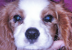
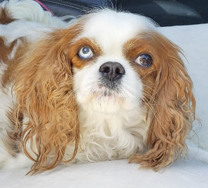
The cavalier King Charles spaniel has more than its fair share of severe diseases afflicting the eye.* A 2008 study of cavaliers conducted by the Canine Eye Registration Foundation showed that an average of 28% of all CKCSs evaluated had eye problems.
* See, American College of Veterinary Ophthalmologists (ACVO) and Ophthalmic Disease in Veterinary Medicine.
They include:
• hereditary cataracts
• corneal dystrophy
• corneal ulcers
• distichiasis
• dry eye syndrome
• entropion
• microphthalmia
• progressive retinal degeneration
• retinal dysplasia
• cherry eye
All are discussed on their own pages on this website. Other hereditary eye disorders, of more minor natures, are listed on this page.
In many instances, the eye disorders CKCSs experience may be attributed to the brachycephalic shape of their heads. Brachycephalic ocular syndrome (BOS) describes these abnormalities attributed to the brachycephalic head shape.
Also, in a November 2016 article, UK ophthalmologists found that the size of the eyes of the cavalier, which is classified by kennel clubs as a small dog, was larger than other small dogs and fit within the eye size of medium sized dogs.
Healthy cavalier puppies have been found to develop mature eyesight between the ages of 30 and 45 days after birth. See this January 2023 article below.
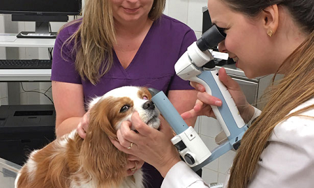
All cavaliers should be examined at least annually by a board certified veterinary ophthalmologist. They are listed on this webpage of the website of the American College of Veterinary Ophthalmologists. See, also our What You Can Do section below for keeping your cavalier's eyes healthy.
© Kip Carter 2021
RETURN TO TOP
List of Eye Disorders
The cavaliers' eye disorders include the following. Click on them to be directed to our articles about them.
- Brachycephalic ocular syndrome
- Cataracts
- Chalazion
- Cherry eye
- Conjunctivitis
- Corneal dystrophy
- Corneal ulcers
- Corneal melting (keratomalacia)
- Conjuntivitis
- Cysts
- Distichiasis
- Dry eye syndrome
- Ectopic cilia
- Ectropion
- Ehlers-Danlos syndrome
- Entropion
- Exophthalmos
- Exotropia
- Eyelid coloboma
- Florida spot keratopathy (FSK)
- Glaucoma
- Heartworm -- Angiostrongylus vasorum in the eye
- Heterochromia
- Horner syndrome
- Hydrocephalus causing blindness
- Hypopyon
- Keratitis
- Keratomycosis
- Lachrymation -- excessive tearing
- Lagophthalmos
- Macroblepharon
- Meibomianitis
- Microphthalmia
- Nystagmus
- Ocular Melanosis
- Optic Neuritis
- Pigmentary Keratitis
- Posterior Lenticonus
- Progressive retinal degeneration
- Proptosis
- Reticulosis
- Retinal detachment
- Retinal dysplasia
- SARDS -- sudden acquired retinal degeneration syndrome
- Squamous cell carcinoma
- Strabismus
- Uveodermatologic syndrome
RETURN TO TOP
Brachycephalic ocular syndrome
Brachycephalic ocular syndrome (BOS) describes eye disorders associated with the conformation of the dog's head known as brachycephalia. "Brachy" means short and "cephalic" means head. The term "brachycephalic" or "brachiocephalic" means short-headed and refers to dogs with a shortened cranium (the bones that house the brain). Most brachycepahic dogs have short muzzles and flat faces and noses which tip back (airorhynchy) and a shorted lower jaw, but not necessarily.
 An
indication of how brachycephalia can affect the eyes of a dog is
"abnormal scleral show", a condition in which the white of the eyeball
is visible above and/or below the iris. In
a
February 2026 article, 73 cavalier King Charles spaniels
were tested for the frequency and severity of Brachycephalic Obstructive
Airway Syndrome (BOAS). The investigators also tested for
"concurrent issues" and found that 36% had
abnormal scleral showing (white part of the eye visible above and/or
below the iris).
An
indication of how brachycephalia can affect the eyes of a dog is
"abnormal scleral show", a condition in which the white of the eyeball
is visible above and/or below the iris. In
a
February 2026 article, 73 cavalier King Charles spaniels
were tested for the frequency and severity of Brachycephalic Obstructive
Airway Syndrome (BOAS). The investigators also tested for
"concurrent issues" and found that 36% had
abnormal scleral showing (white part of the eye visible above and/or
below the iris).
We have two other webpages on this website which discuss cavalier King Charles spaniels having brachycephalic conditions: "Brachycephalic Airway Obstruction Syndrome (BAOS) in the Cavalier King Charles Spaniel" and "The cavalier King Charles spaniel breed is brachycephalic ".
The orbits (eye sockets) of the eyes of many brachycephalic dogs are so shallow that the eye ball protrudes so far forward that it lacks the bony protection which the orbit is intended to provide.
Eye disorders common in CKCSs include Exopthalmos, Exotropia, certain causes of Dry Eye, Lagophthalmos, certain causes of Corneal Ulcers, and Proptosis, all of which are linked and discussed below or elsewhere on this website.
Treating brachycephalic
ocular syndrome (BOS) and the various conditions related to it
include trying to eliminate the abnormalities of the eyes which cause
vision and/or discomfort. Medicine alone usually is insufficent to
resolve these issues. The main surgical option for managing BOS is
called medial canthoplasty. It includes reducing the
size of the palpebral fissure -- the exposed area of
the eyeball between the upper and lower eyelids -- thereby reducing the
eye's exposure and improving the effectiveness of
 blinking and spreading
of tears over the surface and reducing evaporation of tears.
blinking and spreading
of tears over the surface and reducing evaporation of tears.
In this September 2024 article, the authors described the pre-operative and post-operative conditions of a cavalier King Charles spaniel which underwent medial canthoplasty of both eyes. See also this June 2023 article which reviews medial canthoplasty surgeries due to BOS in 271 dogs, including 7 cavaliers.
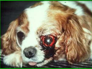
In some cases, when blindness cannot be avoided, the eye(s) may have to be removed, as in the case of Rosie (right) who lost both eyes to glaucoma 7.5 years before this photo was taken at age 14 years. She has continued to live a happy life.
RETURN TO TOP
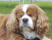
Cataracts
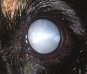 The cavalier King Charles spaniel has a strong breed predisposition
to develop congenital, early-onset juvenile cataracts, which appear by 6
months of age in both eyes and progress to complete cataracts and total
blindness by between ages 2 and 4 years. This form of cataract usually
is combined with other ocular disorders. Older cavaliers also are prone
to develop a non-congenital form of cataract, usually in both eyes,
which also are progressive, and may be expected to form in cavaliers as
old as 7 years of age.
The cavalier King Charles spaniel has a strong breed predisposition
to develop congenital, early-onset juvenile cataracts, which appear by 6
months of age in both eyes and progress to complete cataracts and total
blindness by between ages 2 and 4 years. This form of cataract usually
is combined with other ocular disorders. Older cavaliers also are prone
to develop a non-congenital form of cataract, usually in both eyes,
which also are progressive, and may be expected to form in cavaliers as
old as 7 years of age.
See our Hereditary Cataracts and the Cavalier King Charles Spaniel webpage.
RETURN TO TOP
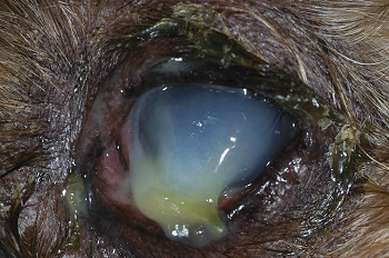
 Cherry Eye
Some cavaliers may develop "prolapsed gland of nictitans", also known as "cherry eye". See our Cherry Eye and the Cavalier King Charles Spaniel webpage.
RETURN TO TOP
Conjunctivitis
Conjunctivitis is a common inflammatory disorder in dogs, and its causes are numerous -- some resulting from other diseases affecting the eyes, and others due to physical injury. The cavalier King Charles spaniel is particularly prone to injuries affecting the conjunctiva of the eye, because of the forward position of the eye in the skulls of many CKCSs.
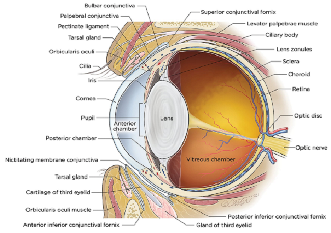
 The conjunctiva is the mucous membrane which covers the back of the
eyelids, as well as the palpebral and bulbar surfaces of the nictitating
membrane which surround the forward surface of the eyeball. (See
diagram above.) The disorder can be either "primary" -- such
as an allergy, immune-mediated inflammation, or an infectious disease --
or "secondary" -- caused by
other eye disorders (e.g.,
keratoconjunctivitis sicca (dry eye),
entropion, ectropion,
distichiasis), allergic
reactions, trauma to the eye (see photo at right of a CKCS with
conjunctivitis resulting from blunt trauma, showing breeding beneath the
conjunctiva), Insect bites and stings, viruses, drug
reactions, infections, parasites, and systemic diseases not intiated at
the eye, including immune disorders.
The conjunctiva is the mucous membrane which covers the back of the
eyelids, as well as the palpebral and bulbar surfaces of the nictitating
membrane which surround the forward surface of the eyeball. (See
diagram above.) The disorder can be either "primary" -- such
as an allergy, immune-mediated inflammation, or an infectious disease --
or "secondary" -- caused by
other eye disorders (e.g.,
keratoconjunctivitis sicca (dry eye),
entropion, ectropion,
distichiasis), allergic
reactions, trauma to the eye (see photo at right of a CKCS with
conjunctivitis resulting from blunt trauma, showing breeding beneath the
conjunctiva), Insect bites and stings, viruses, drug
reactions, infections, parasites, and systemic diseases not intiated at
the eye, including immune disorders.
Typical symptoms of conjunctivitis include:
• Discharges from the eye -- mucus, yellow-green pus, watery
• Fluid build up behind the retina
• Redness (hypermia)
• Swelling
• Discomfort (squinting or rubbing the eye)
• Itchiness
• Ulceration
• Bleeding
Medications control the redness, discharges, pain, and inflammation. They include topical ointments and eye drops, such as corticosteroids or NSAIDs, vasoconsrtictors, antihistamines, and mast cell stabilizers. Other medications vary, depending upon the root cause of the conjunctivitis. If not timely treated, conjunctivitis can lead to more servere eye disorders, including corneal ulcers and corneal melting.
RETURN TO TOP
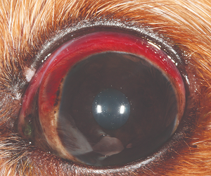
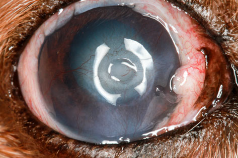
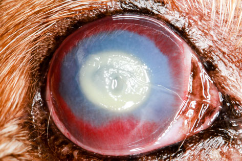
 Corneal Ulcers
Some cavaliers may develop deep abrasions -- ulcers -- in the corneas of their eyes, as a result of their short muzzles and head shapes. See our Corneal Ulcers and the Cavalier King Charles Spaniel webpage.
RETURN TO TOP
 Corneal Melting (Keratomalacia)
Corneal melting (keratomalacia) is a serious condition of the cornea believed to be due to microbial infections which cause inflammation, called infectious keratitis. See our Corneal Melting (Keratomalacia) in Cavalier King Charles Spaniels webpage.
RETURN TO TOP
Cysts
A conjunctival cyst is a cyst on the conjunctiva of the dog's eye. The conjunctiva is the membrane that covers the white part of the eye and lines the inside of the eyelids. Conjunctival cysts are frequently found in the eye socket ("orbit") in dogs. Causes of such cysts include congenital tear duct malformation, production of abnormal secretory material, trauma, or inflammation leading to injury to the walls of the ducts. Many cavaliers suffer from tear duct malformation, which leads to "dry eye" (keratitis sicca or keratoconjunctivitis sicca).
 Surgical
removal of the cyst is the typical remedy. However, treatment choice is
dependent on the size and location of the cyst. Surgery can be
complicated by the proximity of the eye ball, nerves and large vessels,
and the confined space surrounded by bones and ligaments.
Surgical
removal of the cyst is the typical remedy. However, treatment choice is
dependent on the size and location of the cyst. Surgery can be
complicated by the proximity of the eye ball, nerves and large vessels,
and the confined space surrounded by bones and ligaments.
In a June 2021 article, opthalmologist surgeons at Michigan State University reported the successful surgical removal of a conjunctival cyst in the eye of a cavalier (right), using both computed tomography (CT) and digital 3-D surface modeling of the cyst region to plan a minimally invasive approach to remove the cyst while sparing the eye. They concluded that, "Our surgical outcome was excellent with normal function of the nasolacrimal duct and no postoperative complications."
RETURN TO TOP
Distichiasis
 Distichiasis* is the growth of hairs (distichia or cilia) from the
glands of the margin of the upper or lower eyelid, which normally is
hairless. It is considered to be inherited, and the cavalier King
Charles spaniel is predisposed as a breed to this disorder
See our Distichiasis Can Damage Corneas in
Cavaliers webpage.
Distichiasis* is the growth of hairs (distichia or cilia) from the
glands of the margin of the upper or lower eyelid, which normally is
hairless. It is considered to be inherited, and the cavalier King
Charles spaniel is predisposed as a breed to this disorder
See our Distichiasis Can Damage Corneas in
Cavaliers webpage.
RETURN TO TOP
Dry Eye (Keratoconjunctivitis Sicca)
 Many cavaliers suffer from a painful genetic disorder called dry eye
syndrome (keratitis sicca or keratoconjunctivitis sicca -- KCS).
Research studies have shown that cavaliers are more pre-disposed to KCS
-- at a relative risk of 11.5% -- than any other breed.
See our Dry Eye Syndrome and the Cavalier King
Charles Spaniel webpage.
Many cavaliers suffer from a painful genetic disorder called dry eye
syndrome (keratitis sicca or keratoconjunctivitis sicca -- KCS).
Research studies have shown that cavaliers are more pre-disposed to KCS
-- at a relative risk of 11.5% -- than any other breed.
See our Dry Eye Syndrome and the Cavalier King
Charles Spaniel webpage.
RETURN TO TOP
Ectopic Cilia
Ectopic cilia is the growth of hairs (cilia) from the palpebral conjunctiva and rub directly on the cornea, causing severe corneal irritation and in some cases, ulceration. They are similar to distichia but differ as to their source. Both the upper and lower lids can be affected, but the upper lid usually is involved. Cavaliers are reported to be among the breeds predisposed to ectopic cilia. See this March 2020 article and this 2022 book.Treatments for ectopic cilia include destroying the cilia follicles with cautery or cryothermy, or complete surgical excision.
RETURN TO TOP
Ectropion
 Ectopion is the out-turning of the eyelid.The lower lid is more
commonly affected than is the upper lid. It usually is due to the eyelid
being too long for the eye. It usually does not cause irritation, but it
allows exposure of the conjunctiva, which may lead to inflammation.
Also, ectropion may affect normal tear drainage. The photo at right is
of a cavalier with ectropion of both the upper and lower lids, with some
eyelid swelling.
Ectopion is the out-turning of the eyelid.The lower lid is more
commonly affected than is the upper lid. It usually is due to the eyelid
being too long for the eye. It usually does not cause irritation, but it
allows exposure of the conjunctiva, which may lead to inflammation.
Also, ectropion may affect normal tear drainage. The photo at right is
of a cavalier with ectropion of both the upper and lower lids, with some
eyelid swelling.
Medial canthoplasty (MC) is a surgical procedure to correct a variety of eye abnormalities, including ectropion.
RETURN TO TOP
Ehlers-Danlos Syndrome
Ehlers-Danlos syndrome (EDS) is an hereditary connective tissue disease in dogs, humans, and other animals, in which the skin is easily torn. In an October 1987 case in the United Kingdom, a 12-month-old female cavalier King Charles spaniel mix (with border collie) with failing vision also exhibited skin fragility, joint laxity, and ocular signs of bilateral lens luxation, cataract and corneal oedema. It is the first report of ocular signs in EDS in a dog, and joint laxity has been reported only rarely. Neither breed had been implicated previously.
RETURN TO TOP
Entropion
 Entropion is an inward rolling of the eyelid edges, causing the
lashes to rub against the eye's conjunctiva and cornea. Cavaliers have a
relatively high incidence of entropion. It usually develops within a few
months of birth. See our Entropion in the Cavalier King
Charles Spaniel webpage.
Entropion is an inward rolling of the eyelid edges, causing the
lashes to rub against the eye's conjunctiva and cornea. Cavaliers have a
relatively high incidence of entropion. It usually develops within a few
months of birth. See our Entropion in the Cavalier King
Charles Spaniel webpage.
RETURN TO TOP
Exophthalmos
Exophthalmos is the abnormal protrusion of a normal-sized eyeball from its
socket, pushed forward by a space-occupying lesion or object in the
orbit. In cavaliers, exopthalmos has been reportedly due to a variety
of
 causes, including salivary gland disorders known as
sialadenitis and
sialoceles, which is a
swelling of the tissues surrounding the zygomatic gland, which is
located near the eyeballs. Brachycephalia
has been identified as a cause in some CKCSs.
Chiari-like
malformation (CM) also has been associated with exopthalmos.
Masticatory muscle myositis
(MMM) has been associated with exophthalmos in cavaliers.
causes, including salivary gland disorders known as
sialadenitis and
sialoceles, which is a
swelling of the tissues surrounding the zygomatic gland, which is
located near the eyeballs. Brachycephalia
has been identified as a cause in some CKCSs.
Chiari-like
malformation (CM) also has been associated with exopthalmos.
Masticatory muscle myositis
(MMM) has been associated with exophthalmos in cavaliers.
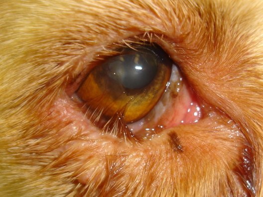
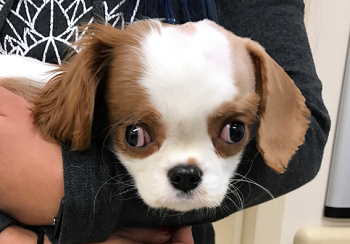
In this March 2025 article, a cavalier was diagnosed with bilateral zygomatic sialadenitis. The zygomatic salivary gland is located just beneath the orbital socket of each eyeball. In this case of an inflamed and enlarged zygomatic gland, a 4-year-old cavalier (see image at right) also had signs of bilateral exophthalmos, dorsolateral globe deviation, third eyelid protrusion, conjunctival hyperaemia, and a superficial corneal ulcer in both eyes due to the corneal exposure, lagophthalmos.
RETURN TO TOP
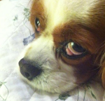
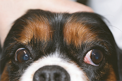
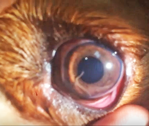
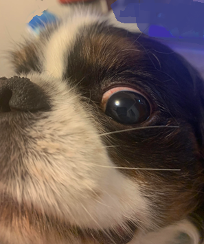
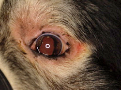
 Exotropia
Exotropia is a condition in which one or both of the dog's eyes deviate outwards. If only one eye is affected, for example the right eye, that eye appears to be looking to the right while the normal eye is looking straight ahead. In the photograph here, the cavalier's right eye has extropia while its left eye is normal.
Exotropia may be caused by Chiari-like malformation (CM) in cavaliers.
Medial canthoplasty (MC) is a surgical procedure to correct a variety of eye abnormalities, including exotropia.
RETURN TO TOP
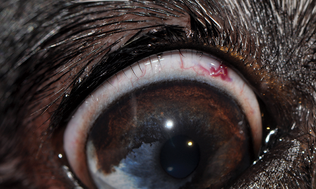
Eyelid Coloboma
 Coloboma
describes a part or entirety of an eyelid which has not properly
developed at birth. It is attributed to the abnormal orientation of the
optic vesicles, which are the fetus' predecessors to the eyes. Eyelid
coloboma is more common in cats, particularly Himalayans, than dogs.
However, it has been reported in the cavalier King Charles spaniel. In
the photo at right, this cavalier has lower eyelid colobomas in both
eyes. Notches in the lower lids are observable, with abnormal hair
growth next to the notches.
Coloboma
describes a part or entirety of an eyelid which has not properly
developed at birth. It is attributed to the abnormal orientation of the
optic vesicles, which are the fetus' predecessors to the eyes. Eyelid
coloboma is more common in cats, particularly Himalayans, than dogs.
However, it has been reported in the cavalier King Charles spaniel. In
the photo at right, this cavalier has lower eyelid colobomas in both
eyes. Notches in the lower lids are observable, with abnormal hair
growth next to the notches.
RETURN TO TOP
Florida Spot Keratopathy (FSK)
Florida spot keratopathy (FSK) (tropical keratopathy, Rice's keratopathy) is a condition of still unknown cause or origin, in which small, round, grayish lesions appear in the corneal stroma, usually with a faint halo surrounding the opaque, circular lesion. Hypotheses of causes range from fire ant bites to environmental conditions such as exposure to ultraviolet lighting. Topical corticosteroids and anti-fungal drugs do not successfully treat FSK, and it usually heals it self over time.
 In
a
December 2024 article, the investigators (O. Pe'er, K. W. Handel, D.
Arad, L. Sebbag, R. Ofri) reported on FSK case studies which 84
FSK-affected dogs, including 6 cavalier King Charles spaniels (7%), were
diagnosed and treated in Israel. They found that most of the dogs had
more than 3 lesions, usually in only one of their eyes. Cavaliers were
the second most prevalent breed in the study, after Labrador retrievers.
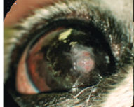
In the image at right, the cavalier has several FSK lesions, following
cataract surgery. The researchers concluded that FSK is a "transient
ocular irritation, with most of them being "non-progressive".
In
a
December 2024 article, the investigators (O. Pe'er, K. W. Handel, D.
Arad, L. Sebbag, R. Ofri) reported on FSK case studies which 84
FSK-affected dogs, including 6 cavalier King Charles spaniels (7%), were
diagnosed and treated in Israel. They found that most of the dogs had
more than 3 lesions, usually in only one of their eyes. Cavaliers were
the second most prevalent breed in the study, after Labrador retrievers.
In the image at right, the cavalier has several FSK lesions, following
cataract surgery. The researchers concluded that FSK is a "transient
ocular irritation, with most of them being "non-progressive".
RETURN TO TOP
Glaucoma
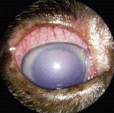 Glaucoma
is a neuro-degenerative disease in which the retinal ganglion cells
begin to die and the optic nerve degenerate. It is one of the leading
causes of blindness in dogs. Canine glaucoma is generally classified as
either primary or secondary. In primary glaucoma, intraocular pressure
(lOP) increases through a reduction in aqueous fluid drainage due to
hereditary abnormality. The most frequent causes of secondary glaucoma
include lens displacement, anterior uveitis (inflammation of the iris
and ciliary body), intraocular neoplasia,
postcataract surgery, and trauma. The cause of the elevated lOP is
generally assumed to be the obstruction of the aqueous humor outflow
pathways. We have found these articles in which cavalier King Charles spaniels
have been
diagnosed with glaucoma:
September 2006,
March 2011,
November 2016.
Glaucoma
is a neuro-degenerative disease in which the retinal ganglion cells
begin to die and the optic nerve degenerate. It is one of the leading
causes of blindness in dogs. Canine glaucoma is generally classified as
either primary or secondary. In primary glaucoma, intraocular pressure
(lOP) increases through a reduction in aqueous fluid drainage due to
hereditary abnormality. The most frequent causes of secondary glaucoma
include lens displacement, anterior uveitis (inflammation of the iris
and ciliary body), intraocular neoplasia,
postcataract surgery, and trauma. The cause of the elevated lOP is
generally assumed to be the obstruction of the aqueous humor outflow
pathways. We have found these articles in which cavalier King Charles spaniels
have been
diagnosed with glaucoma:
September 2006,
March 2011,
November 2016.
RETURN TO TOP
Heartworm -- Angiostrongylus vasorum in the eye
 The French heartworm Angiostrongylus vasorum
(also referred to as a lungworm) is a parasitic
nematode that lives in the pulmonary vessels (in the lung) and the heart of dogs.
A higher occurrence of this nematode parasite has been found in cavaliers than
other breeds, and especially in the cavaliers' eyes as well as the heart
and lung's blood vessels. See
these veterinary reports for details about this
disorder in CKCSs:
June 1994,
2004,
March 2016,
May 2017,
June 2017,
June
2018.
The French heartworm Angiostrongylus vasorum
(also referred to as a lungworm) is a parasitic
nematode that lives in the pulmonary vessels (in the lung) and the heart of dogs.
A higher occurrence of this nematode parasite has been found in cavaliers than
other breeds, and especially in the cavaliers' eyes as well as the heart
and lung's blood vessels. See
these veterinary reports for details about this
disorder in CKCSs:
June 1994,
2004,
March 2016,
May 2017,
June 2017,
June
2018.
RETURN TO TOP
Heterochromia
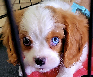 Heterochromia
irides is the scientific term for irises of dogs' eyes having two
different colors, such as one brown eye and one blue eye. Puppies born
with irises of different colors (congenital heterochromia) normally do
not have any vision or other disorders related to it, other than being
more sensitive to light. However, some heterochromia-affected dogs may
have abnormal vision or congenital deafness. Heterochromia is caused by
a lack of the pigment melanin in all or part of the iris of one of the
dog's eyes. Heterochromia is very rare in cavalier King Charles
spaniels, although some lines of CKCSs have it on an hereditary basis.
It is much more common in Australian cattle dogs and shepherds, border
collies, dalmatians, and great Danes.
Heterochromia
irides is the scientific term for irises of dogs' eyes having two
different colors, such as one brown eye and one blue eye. Puppies born
with irises of different colors (congenital heterochromia) normally do
not have any vision or other disorders related to it, other than being
more sensitive to light. However, some heterochromia-affected dogs may
have abnormal vision or congenital deafness. Heterochromia is caused by
a lack of the pigment melanin in all or part of the iris of one of the
dog's eyes. Heterochromia is very rare in cavalier King Charles
spaniels, although some lines of CKCSs have it on an hereditary basis.
It is much more common in Australian cattle dogs and shepherds, border
collies, dalmatians, and great Danes.
RETURN TO TOP
Horner Syndrome
 Claude
Bernard Horner syndrome (Horner's syndrome) is a condition of the dog's
eye in which the ability to control the widening of the eyelid is lost.
The eyelid will appear to droop (ptosis) and the eye's pupil may be constricted
(myosis).
The eye itself may appear to be sunken (enophthalmos), and the third eyelid
(nictitating membrane) may appear reddish and raised. These segments of
the eye are controlled by the part of the dog's involuntary nervous
system called the sympathetic supply to the eye. It is
involved with dilating the pupil and causes the eyelids to open with a
wide-eyed expression, typically in response to stress or excitement.
Horner syndrome is the loss of the sympathetic supply to the eye. In the
photo at right (Copyright © Dr. Clare Rusbridge 2025), the cavalier's
right eye is affected by Horner syndrome, showing a drooping eyelid and
sunken eye.
Claude
Bernard Horner syndrome (Horner's syndrome) is a condition of the dog's
eye in which the ability to control the widening of the eyelid is lost.
The eyelid will appear to droop (ptosis) and the eye's pupil may be constricted
(myosis).
The eye itself may appear to be sunken (enophthalmos), and the third eyelid
(nictitating membrane) may appear reddish and raised. These segments of
the eye are controlled by the part of the dog's involuntary nervous
system called the sympathetic supply to the eye. It is
involved with dilating the pupil and causes the eyelids to open with a
wide-eyed expression, typically in response to stress or excitement.
Horner syndrome is the loss of the sympathetic supply to the eye. In the
photo at right (Copyright © Dr. Clare Rusbridge 2025), the cavalier's
right eye is affected by Horner syndrome, showing a drooping eyelid and
sunken eye.
If only the eye is affected, the disorder is called idiopathic Horner syndrome. However, Horner syndrome may have a wider variety of signs affecting other organs, including the middle ears, brain, spinal cord, and larnyx. Thus, the signs of Horner syndrome can secondary to other disorders, especially infections of those organs.
The cause of idiopathic Horner syndrome remains unknown. If the dog's symptoms are limited to those of Horner syndrome, with no other neurological signs, a diagnosis may be made by giving eye drops of a 1% solution of phenylepineprine, a drug which stimulates the sympathetic nervous system. This treatment may reverse the signs of idiopathic Horner syndrome within 20 minutes. However, this is not to be used as a treatment, as the effect is only temporary, and the side effects of even just 1% of phenylepineprine may sting the eye and cause dilattion of the pupil. Most cases of idiopathic Horner syndrome will cure themselves within 6 months and not likely recur. Thus, the idiopathic version is relatively benign.
For more information, Dr. Clare Rusbridge has published 2 versions of YouTube videos about Horner syndrome. A long version, aimed at veterinarians is linked here. A short version, aimed at dog owners, is linked here.
RETURN TO TOP
Hydrocephalus
 Cavaliers are predisposed to a brain disorder called hydrocephalus,
which may cause blindness.
Hydrocephalus results when excessive amounts of cerebrospinal fluid (CSF) are
produced, either by increased production, or obstruction of its flow, or
the failure of waste-product CSF to be absorbed into the lymphatics and
the bloodstream. Hydrocephalus is the extreme form of enlarged
ventricles (ventriculomegaly) in which
the cavalier suffers clinical symptoms, such as neurological
abnormalities, enlarged skulls, head and neck pain, among a variety of other odd behaviors.
Hydrocephalus creates increased pressure within the cranium and may lead
to degeneration of brain tissue. Blindness occurs if the optic radiation
or occipital cortex is damaged due to hydrocephalus.
Cavaliers are predisposed to a brain disorder called hydrocephalus,
which may cause blindness.
Hydrocephalus results when excessive amounts of cerebrospinal fluid (CSF) are
produced, either by increased production, or obstruction of its flow, or
the failure of waste-product CSF to be absorbed into the lymphatics and
the bloodstream. Hydrocephalus is the extreme form of enlarged
ventricles (ventriculomegaly) in which
the cavalier suffers clinical symptoms, such as neurological
abnormalities, enlarged skulls, head and neck pain, among a variety of other odd behaviors.
Hydrocephalus creates increased pressure within the cranium and may lead
to degeneration of brain tissue. Blindness occurs if the optic radiation
or occipital cortex is damaged due to hydrocephalus.
See our Hydrocephalus webpage for more information about this disorder.
RETURN TO TOP
Hypopyon
Hypopyon is the presence of inflammatory cells, especially leukocytes, in the rear chamber of the eye. Hypopyon can lead to secondary glaucoma. Hypopyon is typically treated with anti-inflammatory medications, as it is an accumulation of inflammatory cells. Corneal ulcers, corneal abscesses, uveitis (inflammation of the uvea), and systemic illnesses commonly cause hypopyon.
RETURN TO TOP
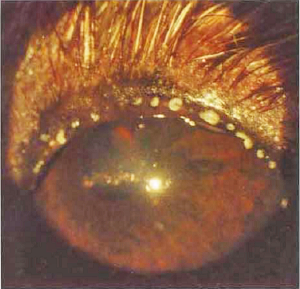
Keratitis
Keratitis is an inflammation of the cornea, the anterior part of the eye which covers the pupil. Corneal inflammations can be caused by bacteria, fungus or virus - but whatever the agent the cornea is going to present some quite nasty symptoms. Keratitis sicca is also known as "dry eye syndrome", which we cover separately at its own webpage.
We have found two veterinary journal articles
describing the diagnoses and treatments of cavaliers with keratitis
infections. Both of them included surgeries.
 In this
September 2015 article, a
cavalier was among five dogs with keratomycosis, an infection in the
cornea caused by a fungus. Due to the severity of the infection, a
keratectomy (corneal surgery) and conjunctival graft surgery were
successfully performed. See the before (left) and after (right)
photos of this cavalier's eye.
In this
September 2015 article, a
cavalier was among five dogs with keratomycosis, an infection in the
cornea caused by a fungus. Due to the severity of the infection, a
keratectomy (corneal surgery) and conjunctival graft surgery were
successfully performed. See the before (left) and after (right)
photos of this cavalier's eye.
In this June 2016 article, a cavalier was one of six dogs and two cats studied. The CKCS had infectious crystalline keratopathy (ICK) in its cornea. An anterior lamellar keratectomy and a corneoconjunctival transposition surgical procedure were successfully performed.
RETURN TO TOP
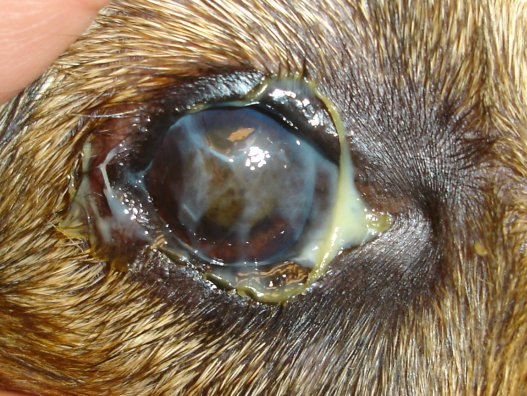
Keratomycosis
In this September 2015 article, a cavalier was diagnosed with keratomycosis, an infection in the cornea caused by a fungus. There also was a corneal ulceration with surrounding keratomalacia, corneal white cell infiltrate, and mild hypopyon. Due to the severity of the infection, a keratectomy (corneal surgery) and conjunctival graft surgery were successfully performed.
RETURN TO TOP
Lachrymation -- excessive tearing
 Lachrymation, the excessive secretion of tears, often is combined
with epiphora, the inadequate drainage of tears.
In cavaliers,
this may be attributed to a congenital condition involving either the
lack of an opening in the tear duct to allow for the drainage of tears,
or due to an inadequately sized opening. The opening is called the
lacrimal punctum. There are two of them in each eye --
a superior and an inferior. An absence of one or both of them is called imperforate
lactimal punctum, and an inadequately sized one is called a
micro-lachrymal punctal. When one or both are blocked,
the tears that lubricate the eye have no place to drain other than over
the rim of the eye and down the face. Symptoms are tear staining of the
hair around the eye.
Lachrymation, the excessive secretion of tears, often is combined
with epiphora, the inadequate drainage of tears.
In cavaliers,
this may be attributed to a congenital condition involving either the
lack of an opening in the tear duct to allow for the drainage of tears,
or due to an inadequately sized opening. The opening is called the
lacrimal punctum. There are two of them in each eye --
a superior and an inferior. An absence of one or both of them is called imperforate
lactimal punctum, and an inadequately sized one is called a
micro-lachrymal punctal. When one or both are blocked,
the tears that lubricate the eye have no place to drain other than over
the rim of the eye and down the face. Symptoms are tear staining of the
hair around the eye.
The cavalier King Charles spaniel is not reported to be pre-disposed to imperforate or micro-lachrymal puncta, but in a 1979 article, British ophthalmologist Keith Barnett reported the case of a cavalier diagnosed with this disorder, associated with microphthalmos. See this website for more details of this condition in general.
RETURN TO TOP
Lagophthalmos
 Lagophthalmos -- the inability to fully close the eyelids -- can
result in inadequate blinking, causing dry, irritated eyes and possible
scarring. The result is called exposure keratitis. See this
May 2015 article. A surgical procedure called tarsorrhaphy,
in which the eyelids are partially sewn together to narrow the eyelid
opening, may have to be performed.
Lagophthalmos -- the inability to fully close the eyelids -- can
result in inadequate blinking, causing dry, irritated eyes and possible
scarring. The result is called exposure keratitis. See this
May 2015 article. A surgical procedure called tarsorrhaphy,
in which the eyelids are partially sewn together to narrow the eyelid
opening, may have to be performed.
In this March 2025 article, a cavalier was diagnosed with bilateral zygomatic sialadenitis. The zygomatic salivary gland is located just beneath the orbital socket of each eyeball. In this case of an inflamed and enlarged zygomatic gland, a 4-year-old cavalier (see image at right) also had signs of bilateral exophthalmos, dorsolateral globe deviation, third eyelid protrusion, conjunctival hyperaemia, and a superficial corneal ulcer in both eyes due to the corneal exposure, lagophthalmos.
Medial canthoplasty (MC) is another surgical procedure to correct a variety of eye abnormalities, including lagophthalmos.
RETURN TO TOP
Macroblepharon
 Macroblepharon
is a condition in which there dog's eye protrudes due to excessive
length of the eyelid. It reportedly is common among brachycephalic
breeds, including the cavalier. A usual sign of this disorder is the
white of the eye (sclera) being more visible, along with the eyeball
bulging forward. It can be the cause of
corneal ulcers, due to the excessive exposure.
Medial canthoplasty, a surgical procedure described below, is often
performed to resolve this disorder. It involves reducing the
eyelid length and preventing abrasion of the eye's surface.
Macroblepharon
is a condition in which there dog's eye protrudes due to excessive
length of the eyelid. It reportedly is common among brachycephalic
breeds, including the cavalier. A usual sign of this disorder is the
white of the eye (sclera) being more visible, along with the eyeball
bulging forward. It can be the cause of
corneal ulcers, due to the excessive exposure.
Medial canthoplasty, a surgical procedure described below, is often
performed to resolve this disorder. It involves reducing the
eyelid length and preventing abrasion of the eye's surface.
In this photo of a cavalier's left eye, it has macroblepharon, along with dry eye and corneal ulceration.
RETURN TO TOP
Meibomianitis
 Meibomianitis is an inflammation of the meibomian glands, which are
sebaceous glands located in the eyelid and provide the eye with an
oily substance which lubricates the tear film covering the cornea.
If one of the glands becomes blocked, it will retain the oil and swell.
An enlarged meibomian gland also is called a chalazion.
Meibomianitis is an inflammation of the meibomian glands, which are
sebaceous glands located in the eyelid and provide the eye with an
oily substance which lubricates the tear film covering the cornea.
If one of the glands becomes blocked, it will retain the oil and swell.
An enlarged meibomian gland also is called a chalazion.
Meibomianitis may also be diagnosed with inflammation of the eyelid and/or conjuctivitis. The cavalier in the photo at the right has meibomianitis,. The whiteish spots are secretions from the meibomian glands.
RETURN TO TOP
 Microphthalmia
Microphthalmia describes a condition in which one or both of the dog's eyes is abnormally small, resulting in restricted vision and possible blindness. It is a congenital and often inherited defect which is particularly common in the cavalier. See our Microphthalmia in Cavalier King Charles Spaniels webpage.
RETURN TO TOP
Nystagmus
Nystagmus is the uncontrolled movement of the eyes, usually from side to side, or in a circular pattern. It may be a symptom resulting from such disorders as retinal dysplasia, cerebellar infarcts (strokes), icterus (jaundice), forms of vestibular syndrome, and primary secretory otitis media (PSOM).
RETURN TO TOP
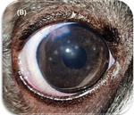
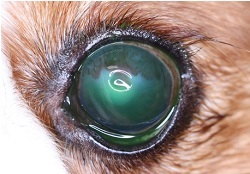
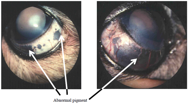
Ocular Melanosis
Ocular melanosis is a congenitial eye condition that starts with increased black or dark brown pigmentation in the iris of the eye. It progresses onto the sclera (white) of the eye, and eventually in most cases, to increased pressure within the eye, resulting in glaucoma. (See comparative photos below.) The condition cannot be treated successfully, and typically causes pain and loss of vision. It has been found to be most common in Cairn terriers.
In a January 2024 article, University of Wisconsin researchers examined the medical records of dogs diagnosed with ocular melanosis at the Comparative Ocular Pathology Laboratory of Wisconsin over a ten year period. In this current study, cavalier King Charles spaniels were found to be at a "relative risk" of 1.7. However, the researchers report that due to the small number of diagnosed cases of cavaliers, only 8, they may be able to conclude ony that the CKCS "may also be at risk".
RETURN TO TOP
Optic Neuritis
Optic neuritis is a rare but serious condition that can result in acute blindness or visual deficits in one or both eyes. It has been reported in cavalier King Charles spaniels in at least three peer-reviewed veterinary publications. See this January 1981 article and this February 2004 article. and this September 2020 article.
Optic neuritis describes various diseases of the optic nerve that cause primary demyelination and appear as a sudden visual field defect or total loss of vision in one or both eyes. It is associated with increased intracranial pressure and is primarily a mechanical, not a vascular phenomenon. Optic nerve fibers are compressed by elevated cerebrospinal fluid (CSF) pressure in the optic nerve, resulting in swelling. Optic nerve atrophy and vision loss usually occur after a few weeks, due to nerve fiber attrition.
For more information, see this March 2018 article.
RETURN TO TOP
Pigmentary Keratitis
 Pigmentary keratitisis a brownish-black discoloration patches in the
cornea and visible on the eye's surface caused by the deposition of
pigmented melanin granules. It is not a disorder in and of itself;
instead, it is a result of some other, underlying eye or eyelid disorder
which has caused chronic irritation or inflammation. The inflammation in
turn causes melanin granules to be deposited in the cornea.Typical
causations are dry eye (keratoconjunctivitis
sicca), corneal ulcers,
entropion, ectropion,
or other eyelid or eyelash disorders.
Pigmentary keratitisis a brownish-black discoloration patches in the
cornea and visible on the eye's surface caused by the deposition of
pigmented melanin granules. It is not a disorder in and of itself;
instead, it is a result of some other, underlying eye or eyelid disorder
which has caused chronic irritation or inflammation. The inflammation in
turn causes melanin granules to be deposited in the cornea.Typical
causations are dry eye (keratoconjunctivitis
sicca), corneal ulcers,
entropion, ectropion,
or other eyelid or eyelash disorders.
In a November 2024 article, researchers found that all cavaliers in the study had pigmentary keratitis, making it the most prevalent pre-existing condition in the study.
RETURN TO TOP
Posterior Lenticonus
Lenticonus is a congenital deformity in dogs, in which an area of the lens surface protrudes in a cone-like shape bulge. Posterior lenticonus identifies the location of the protrusion, the back of the lens. It is considered rarely seen in dogs.
Posterior lenticonus has been reported in the cavalier King Charles spaniel along with a few other breeds. The condtion may enlarge with age, developing into a cataract and even rupturing the lens. As it progresses, the lenticonus may be painful and vision become opaque. See this 2022 book.
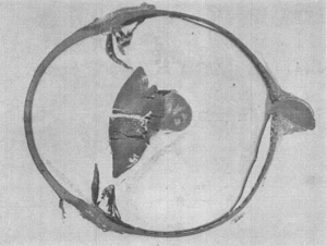 In
this
November 1984 article, 3 cavaliers in Sweden were diagnosed with
this disorder, and one other CKCS was suspected of it. In all cases,
additional optical disorders -- cataracts and microphthalmia -- also
were observed. In one of these cases, a 2-month-old cavalier puppy had a
cone-like protrusion of the posterior surface of the lens ("lenticular
surface") of the right eye. In the other eye, they found a larger
protrusion of the posterior lens. Three months later, the puppy became
blind and was in great pain. The lens bulges had progressed, and the
left eye had a complete cataract. (The image above shows a gross
section of the puppy's left eye, with bulging of the lens material from
the posterior region of the lens into the vitreous.)
In
this
November 1984 article, 3 cavaliers in Sweden were diagnosed with
this disorder, and one other CKCS was suspected of it. In all cases,
additional optical disorders -- cataracts and microphthalmia -- also
were observed. In one of these cases, a 2-month-old cavalier puppy had a
cone-like protrusion of the posterior surface of the lens ("lenticular
surface") of the right eye. In the other eye, they found a larger
protrusion of the posterior lens. Three months later, the puppy became
blind and was in great pain. The lens bulges had progressed, and the
left eye had a complete cataract. (The image above shows a gross
section of the puppy's left eye, with bulging of the lens material from
the posterior region of the lens into the vitreous.)
The puppy was put down, and its eyes were preserved for further investigation. They found that the posterior lens capsule had ruptured in both eyes. Clusters of white blood cells called macrophages had surrounded the ruptured area and the lining of the iris. A review of pedigrees of the 3 affected cavaliers led the investigators to conclude that posterior lenticonus is heritable in the breed.
RETURN TO TOP
Progressive Retinal Atrophy
Progressive retinal atrophy (PRA), also known as progressive retinal degeneration (PRD), is a disease which causes blindness and has "increased incidence" and is "presumed inherited" in the cavalier. See our Progressive Retinal Atrophy in Cavaliers webpage.
RETURN TO TOP
 Proptosis
Proptosis is the protrusion of an eyeball, at least partially out of its socket (orbit). It most commonly is caused by a traumatic event, such as a blunt-force injury or trauma from physical restraint. The eyelid becomes trapped behind the eyeball, preventing the eyeball from returning to its normal position. Cavaliers, due to their brachycephalic skull structure, may be susceptible to proptosis due to the shallowness of their eye orbits. This disorder always is very serious and requires immediate emergency treatment.
RETURN TO TOP
Reticulosis
Reticulosis is a cellular reaction to inflammation that mainly affects the central nervous system. Reticulosis of the eye is an inflammatory condition which has infiltrated the optic nerves. It is considered a form of optic neuritis, which is described above. In this January 1981 artcle, a cavalier King Charles spaniel was diagnosed with reticulosis after having been blind for two days and regained its sight after treatment with prednisolone and chloramphenicol for five days. The dog lost its vision again two weeks after six weeks of prednisolone treatment. Steroid treatment was ineffective thereafter, and due to other symptoms of the central nervous system, the dog was euthanized. Upon necropsy of the eyes, the optic nerves were found to be swollen, and the left optic nerve was twice as thick as the right one. The entire spinal cord was inveloped with perivascular infilltrates in the cells. The diagnosis therefore was inflammatory reticulosis of the central nervous system and the eyes.
RETURN TO TOP
Retinal Detachment
 Retinal
detachment is the separation of the rtina between its pigmented
epithelium (RPE) and its sensory layer. The causes are varied any can
include trauma causing a tear that allows fluid to enter between the
layers of the retina, or the formation of new growth (neoplasia), or
inflammation, or vascular disease, such as hypertension, or degenerative
changes, or retinal dysplasia, or
some types of cataracts, among others.
Severe cases of retinal detachment often result in reduced vision or
blindness. Cavaliers are not immune to this condtion, as indicated by
the photo at right, of a cavalier's right eye with a detached retina.
Retinal
detachment is the separation of the rtina between its pigmented
epithelium (RPE) and its sensory layer. The causes are varied any can
include trauma causing a tear that allows fluid to enter between the
layers of the retina, or the formation of new growth (neoplasia), or
inflammation, or vascular disease, such as hypertension, or degenerative
changes, or retinal dysplasia, or
some types of cataracts, among others.
Severe cases of retinal detachment often result in reduced vision or
blindness. Cavaliers are not immune to this condtion, as indicated by
the photo at right, of a cavalier's right eye with a detached retina.
RETURN TO TOP
Retinal Dysplasia
Retinal dysplasia is a congenital malformation of the retina. It occurs when the two layers of the retina do not form together properly. See our Retinal Dysplasia in Cavalier King Charles Spaniels webpage.
RETURN TO TOP
SARDS -- sudden acquired retinal degeneration syndrome
A cause of late-onset retinal dysplasia is a condition known as sudden acquired retinal degeneration syndrome (SARDS), See our SARDS section of our Retinal Dysplasia in Cavalier King Charles Spaniels webpage.
RETURN TO TOP
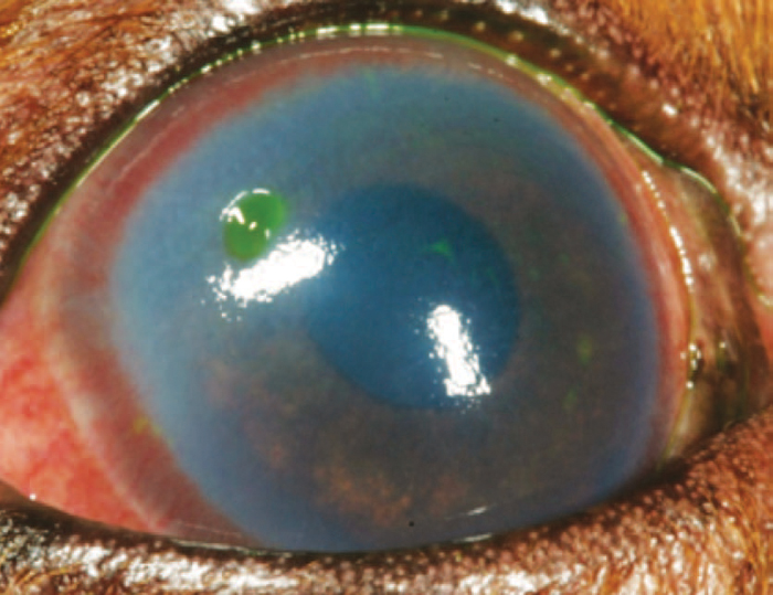
Squamous Cell Carcinoma
 Squamous cell carcinoma (SCC) is a common skin cancer in humans and
dogs, which may include the eyelid. In the cavalier King Charles spaniel
and a limited number of other breeds, however, it can attack the cornea.
It is caused by an excessive production of squamous cells in the outer
layer of skin. In the eye, SCC has been described as a pink, elevated,
and cauliflower-like mass on the surface of the cornea (right). SCC must be
identified and treated promptly, as it can be an aggressive cancer which can
result in blindness. In a
June 2009 article, Dr. Kerry Ketring wrote
that:
Squamous cell carcinoma (SCC) is a common skin cancer in humans and
dogs, which may include the eyelid. In the cavalier King Charles spaniel
and a limited number of other breeds, however, it can attack the cornea.
It is caused by an excessive production of squamous cells in the outer
layer of skin. In the eye, SCC has been described as a pink, elevated,
and cauliflower-like mass on the surface of the cornea (right). SCC must be
identified and treated promptly, as it can be an aggressive cancer which can
result in blindness. In a
June 2009 article, Dr. Kerry Ketring wrote
that:
"This isolated mass is most commonly seen in older Cavalier King Charles spaniels, pugs, and Shih Tzus. These tumors can be removed with care by a stromal keratectomy. Laser therapy, B-radiation, and cryotherapy have been recommended following the keratectomy."
In both a 2008 study and a May 2011 study of this cancer in the cornea, which included three CKCSs, the authors found that it was related to chronic dry eye, which is a very common disorder in this breed. This form of carcinoma also is known to develop within the mouths and throats of cavaliers.
In a November 2024 article of the recurrence rate of SCC in 16 dogs, 3 of them, the second most common breed, were cavaliers.
RETURN TO TOP
Strabismus
 Strabismus
defines the condition in which one of the dog's eyes cannot focus with
the other on an object because of an imbalance of the eye muscles.
Strabismus
defines the condition in which one of the dog's eyes cannot focus with
the other on an object because of an imbalance of the eye muscles.
Dr. David Williams reported of a case of a cavalier in which "it was impossible to move the globe medially and there was a lateral post-traumatic restritive strabismus which the owners chose not to treat as the dog was not impaired by the problem." (See the photograph at right.)
In a July 2014 article, Drs. Rusbridge, Knowler, and others reported on cavaliers with a combination of lateral strabismus along with other eye conformational abnormalities (exophthalmos and exotropia) and their association with Chiari-like malformation.
RETURN TO TOP
Uveodermatologic syndrome
 Canine
uveodermatologic syndrome* (CUDS) is an
autoimmune disease in which the dog's immune system forms antibodies
against its pigment cells (melanocytes) and its light-sensing cells in
its eyes' retinas. It can cause redness, inflammation of the uvea (uveitis)
and pain in the eyes, and also affects the dog's skin, causing loss of
pigmentation (vitiligo), particularly on the face, hind end, and paws,
and whitening of the dog's hair (poliosis). Exposure to sunlight may
worsen the conditions. Blindness is a likely result if treatment is not
rapid and thorough. Suppressing the immune system with glucocorticoids
or cyclosporine is the usual approach
to relieve the inflammation and pain and to slow progression of the
disease. Some researchers suspect that the underlying cause is a virus.
Canine
uveodermatologic syndrome* (CUDS) is an
autoimmune disease in which the dog's immune system forms antibodies
against its pigment cells (melanocytes) and its light-sensing cells in
its eyes' retinas. It can cause redness, inflammation of the uvea (uveitis)
and pain in the eyes, and also affects the dog's skin, causing loss of
pigmentation (vitiligo), particularly on the face, hind end, and paws,
and whitening of the dog's hair (poliosis). Exposure to sunlight may
worsen the conditions. Blindness is a likely result if treatment is not
rapid and thorough. Suppressing the immune system with glucocorticoids
or cyclosporine is the usual approach
to relieve the inflammation and pain and to slow progression of the
disease. Some researchers suspect that the underlying cause is a virus.
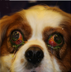
In this October 2024 article, a cavalier was diagnosed with both uveodermatological syndrome and alopecia areata, which is a hair-loss condtion also due to immune system disfunctions. The 1.5 year old female tricolor previously had been diagnosed with bilateral uveitis and keratoconjunctivitis sicca, resulting in bilateral glaucoma. Owing to poor response, removal of both eyes was elected. Histopathological evaluation of ocular globes confirmed the presence of lympho-histiocytic uveitis and the potential of CUDS was suggested.
* Uveodermatologic syndrome is related to a condition in humans called Vogt-Koyanagi-Harada-like syndrome.
RETURN TO TOP
Diagnosis
 Each disorder of the eyes may call for a different manner of
diagnosis and certainly a different form of treatment, but the initial
diagnosis steps, which should be performed by the cavalier's
veterinarian at every visit, is fairly standard. There are two initial
steps: (1) Schirmer tear
testing (STT) and (2)
fluorescein staining (right).
Each disorder of the eyes may call for a different manner of
diagnosis and certainly a different form of treatment, but the initial
diagnosis steps, which should be performed by the cavalier's
veterinarian at every visit, is fairly standard. There are two initial
steps: (1) Schirmer tear
testing (STT) and (2)
fluorescein staining (right).
When a disorder is suspected, the dog also should receive a thorough ophthalmic examination, including evaluation of the menace response, dazzle reflex, pupillary light reflex, slit-lamp examination, Schirmer Tear Test I, and rebound tonometry.
RETURN TO TOP
Treatment
The treatment will vary, depending upon the particular disorder and its severity. It may extend from daily eye drops to major surgery.
Treating brachycephalic
ocular syndrome (BOS) and the various conditions related to it
include trying to eliminate the abnormalities of the eyes which cause
vision and/or discomfort. Medcine alone usually is insufficent to
resolve these issues. The main surgical option for managing BOS is
called medial canthoplasty. It includes reducing the
size of the palpebral fissure -- the exposed area of
the eyeball between the upper and lower eyelids -- thereby reducing the
eye's exposure and improving the effectiveness of
blinking and spreading
of tears over the surface and reducing evaporation of tears.
In this September 2024 article, the authors described the pre-operative and post-operative conditions of a cavalier King Charles spaniel which underwent medial canthoplasty of both eyes. See also this June 2023 article which reviews medial canthoplasty surgeries due to BOS in 271 dogs, including 7 cavaliers.
In some cases, when blindness cannot be avoided, the eye(s) may have to be removed, as in the case of Rosie (right) who lost both eyes to glaucoma 7.5 years before this photo was taken at age 14 years. She has continued to live a happy life.
RETURN TO TOP
Related Links
RETURN TO TOP
 What You Can Do:
Annual Check-Ups
All CKCSs should be examined at least annually by a board certified veterinary ophthalmologist. They are listed on the website of the ACVO.
Daily Wipes
Consider wiping beneath your cavalier's eyes daily to remove drainage from the tear ducts which may attract or retain bacteria. Pre-moistened OptixCare wipes are designed for that purpose.
Rinsing
If your cavalier appears to be wincing one of its eyes, you may safely rinse the eye with a sterile saline solution.
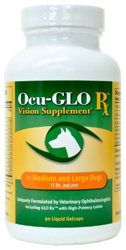 Eye
Supplement
Eye
Supplement
Ocu-GLO Rx is a nutraceutical containing several natural antioxidants in a combination blend formulated specifically for canine eye health. Many veterinary ophthalmologists recommend this product to maintain healthy eyes. Even if your dog has not been diagnosed with a vision disorder, antioxidants contained in Ocu-GLO Rx are considered helpful in keeping dogs' eyes healthy.
Blind Cavaliers
Blind cavailers can almost 'see' with their sense of smell. In a
July 2022 article, researchers
examined the olfactory systems of 23 laboratory dogs to determine the
extent of tracts connecting the dogs;
olfactory bulbs with regions of
their brains. They proceeded upon the theory of an integration of smell
and vision in dog cognition. They used diffusion tensor imaging (DTT)
MRI and a process for dissecting brain fibers, known as the Klingler
method. They report finding five tracts of "white matter" connecting the
olfactory bulb to the cortex of the brain. The tracts are: (1)
olfactory-occipital tract (OOT); (2) olfactory-cortical spinal tract
(OCST); (3) olfactory-limbic tract (OLT); (4) olfactory-piriform tract
(OPT); and (5) olfactory-entorhinal tract (OET). They concluded
that:
"These findings show the dog's olfactory system is integrated with many different parts of the brain. The OOT [occipital cortex] connection is of particular interest due to its size and relevance to understanding canine cognition. The presence of an olfactory-occipital 'information highway' provides structural evidenc for the theorized olfactory-vision integration proposed in canine cognitive research."
Researcher Dr. Johnson explained about seeing with their noses, in an interview about her article:
"We've never seen this connection between the nose and the occipital lobe, functionally the visual cortex in dogs, in any species. When we walk into a room, we primarily use our vision to work out where the door is, who's in the room, where the table is. Whereas in dogs, this study shows that olfaction is really integrated with vision in terms of how they learn about their environment and orient themselves in it. They can still play fetch and navigate their surroundings much better than humans with the same condition. Knowing there's that information freeway going between those two areas could be hugely comforting to owners of dogs with incurable eye diseases. Veterinarians have long wondered how dogs with complete blindness have navigated so well in their environment, even in foreign and new environments. The olfactory connection we identified gives us an answer to this, and shows that they are less dependent on their eyes alone and likely use olfactory information to help navigate their world."
So, do not deprive your blind cavalier of opportunities to be out on walks or carriage rides and smell their environments.
RETURN TO TOP
Research News:
March 2025:
Inflamed zygomatic salivary glands cause multiple eye disorders
to a cavalier.
In
a
March 2025 article, UK researchers (A. E. Enache, S. Maini, M.
Pivetta, E. Jeanes, L. Fleming, C. Hartley, R. Tetas Pont) reported 20
cases of bilateral zygomatic sialadenitis, the inflammation and swelling
of the zygomatic salivary glands. These glands are located just beneath
the eyeballs. Of the 20, most patients were Labrador retrievers and 2
were cavalier King Charles spaniels. In Case #9, a 4-year-old CKCS
(right) had both of its zygomatic glands inflammed and enlarged.
This swelling of the glands caused several eye disorders, including
exophthalmos, dorsolateral globe deviation, third eyelid protrusion,
conjunctival hyperaemia, and a superficial corneal ulcer in both eyes
due to lagophthalmos -- the inability to close the eyelids. The dog was
treated with the anti-microbial amoxicillin for 2 weeks, prednisolone
for a month, omeprazole for 1 week, methadone, chloramphenicol, and
celluvisc. Signs began to improve in the first 24 hours. After 22 days,
the only remaining condition was prolapse of a third eyelid gland.
December 2024:
Cavaliers ranked second among breeds diagnosed with Florida Spot
Keratopathy among 84 dogs.
 Florida spot keratopathy (FSK) (tropical keratopathy, Rice's
keratopathy) is a condition of still unknown cause or origin, in which
small, round, grayish lesions appear in the corneal stroma, usually with
a faint halo surrounding the opaque, circular lesion. Hypotheses of
causes range from fire ant bites to environmental conditions such as
exposure to ultraviolet lighting. Topical corticosteroids and
anti-fungal drugs do not successfully treat FSK, and it usually heals it
self over time.
Florida spot keratopathy (FSK) (tropical keratopathy, Rice's
keratopathy) is a condition of still unknown cause or origin, in which
small, round, grayish lesions appear in the corneal stroma, usually with
a faint halo surrounding the opaque, circular lesion. Hypotheses of
causes range from fire ant bites to environmental conditions such as
exposure to ultraviolet lighting. Topical corticosteroids and
anti-fungal drugs do not successfully treat FSK, and it usually heals it
self over time.
In a December 2024 article, the investigators (O. Pe'er, K. W. Handel, D. Arad, L. Sebbag, Ron Ofri [right]) reported on FSK case studies which 84 FSK-affected dogs, including 6 cavalier King Charles spaniels (7%), were diagnosed and treated in Israel. They found that most of the dogs had more than 3 lesions, usually in only one of their eyes. Cavaliers were the second most prevalent breed in the study, after Labrador retrievers. The researchers concluded that FSK is a "transient ocular irritation, with most of them being "non-progressive".
October 2024:
Cavalier is diagnosed with uveodermatological syndrome and
alopecia areata.
 In
an
October 2024 article, a team of American clinicans (Barbara G.
McMahill [right], Sophie Gilbert, Jamie Haddad, Janelle Novak,
Maria Shank, and Verena K. Affolter) report a case study of a year old
female cavalier King Charles spaniel they diagnosed with both
uveodermatological syndrome and alopecia areata. Uveodermatologic
syndrome is an autoimmune disease in which the dog's immune system forms
antibodies against its pigment cells (melanocytes) and its light-sensing
cells in its eyes' retinas. It can cause redness, inflammation
(uveitis) and pain in the eyes, and also affects the dog's skin,
causing loss of pigmentation (vitiligo), particularly on the face, hind
end. and paws, and whitening of the dog's hair (poliosis). Exposure to
sunlight may worsen the conditions. Blindness is a likely result if
treatment is not rapid and thorough. Alopecia areata is a hair-loss
condtion also due to immune system disfunctions. They describe the dog's
signs, diagnosis, treatment, and clinical follow-up.
In
an
October 2024 article, a team of American clinicans (Barbara G.
McMahill [right], Sophie Gilbert, Jamie Haddad, Janelle Novak,
Maria Shank, and Verena K. Affolter) report a case study of a year old
female cavalier King Charles spaniel they diagnosed with both
uveodermatological syndrome and alopecia areata. Uveodermatologic
syndrome is an autoimmune disease in which the dog's immune system forms
antibodies against its pigment cells (melanocytes) and its light-sensing
cells in its eyes' retinas. It can cause redness, inflammation
(uveitis) and pain in the eyes, and also affects the dog's skin,
causing loss of pigmentation (vitiligo), particularly on the face, hind
end. and paws, and whitening of the dog's hair (poliosis). Exposure to
sunlight may worsen the conditions. Blindness is a likely result if
treatment is not rapid and thorough. Alopecia areata is a hair-loss
condtion also due to immune system disfunctions. They describe the dog's
signs, diagnosis, treatment, and clinical follow-up.
January 2024:
Cavaliers may be at risk for ocular melanosis.
 In
a
January 2024 article, University of Wisconsin researchers (J. Seth
Eaton [right], Sanskruti S. Potnis, Alexis Cavanaugh, Cody A.
Davis, Leandro B. C. Teixeira, Gillian C. Shaw) examined the medical
records of dogs diagnosed with ocular melanosis at the Comparative
Ocular Pathology Laboratory of Wisconsin over a ten year period. Ocular
melanosis is a congenitial eye condition that starts with increased
black or dark brown pigmentation in the iris of the eye. It progresses
onto the sclera (white) of the eye, and eventually in most cases, to
increased pressure within the eye, resulting in glaucoma. (See
comparative photos below.) The condition cannot be treated
successfully, and typically causes pain and loss of vision. It has been
found to be most common in Cairn terriers. In this current study,
cavalier King Charles spaniels were found to be at a "relative risk" of
1.7. However, the researchers report that due to the small number of
diagnosed cases of cavaliers, only 8, they may be able to conclude ony
that the CKCS "may also be at risk".
In
a
January 2024 article, University of Wisconsin researchers (J. Seth
Eaton [right], Sanskruti S. Potnis, Alexis Cavanaugh, Cody A.
Davis, Leandro B. C. Teixeira, Gillian C. Shaw) examined the medical
records of dogs diagnosed with ocular melanosis at the Comparative
Ocular Pathology Laboratory of Wisconsin over a ten year period. Ocular
melanosis is a congenitial eye condition that starts with increased
black or dark brown pigmentation in the iris of the eye. It progresses
onto the sclera (white) of the eye, and eventually in most cases, to
increased pressure within the eye, resulting in glaucoma. (See
comparative photos below.) The condition cannot be treated
successfully, and typically causes pain and loss of vision. It has been
found to be most common in Cairn terriers. In this current study,
cavalier King Charles spaniels were found to be at a "relative risk" of
1.7. However, the researchers report that due to the small number of
diagnosed cases of cavaliers, only 8, they may be able to conclude ony
that the CKCS "may also be at risk".
June 2023:
Cavaliers ranked fourth among breeds in UK recommended for
eyelid surgery due to brachycephalic conditions.
 In
a
June 2023 article, UK researchers (Amy L. M. M. Andrews [right],
Katie L. Youngman, Rowena M. A. Packer, Dan G. O'Neill, Christiane
Kafarnik) reviewed veterinary records of 271 brachycephalic dogs with
eye conditions severe enough to be recommended to have eyelid surgeries
(medial canthoplasty) between 2016 and 2021. Ranked first was the pug
(71.6%), followed by the shih tzu (17.7%), French bulldog (5.5%), and
cavalier King Charles spaniel (2.6%). Medial canthoplasty (MC) is a
surgical procedure to correct a variety of eye abnormalities, including
entropion, extropia, exposure dry eye, lagophthalmos, and the risk of
painful corneal ulceration. Among the 81 cavaliers examined at the Queen
Mother Hospital for Animals for ophthalmological conditions, 7 of them
(8.6%) were recommended to have this form of surgery during the
2016-2021 period.
In
a
June 2023 article, UK researchers (Amy L. M. M. Andrews [right],
Katie L. Youngman, Rowena M. A. Packer, Dan G. O'Neill, Christiane
Kafarnik) reviewed veterinary records of 271 brachycephalic dogs with
eye conditions severe enough to be recommended to have eyelid surgeries
(medial canthoplasty) between 2016 and 2021. Ranked first was the pug
(71.6%), followed by the shih tzu (17.7%), French bulldog (5.5%), and
cavalier King Charles spaniel (2.6%). Medial canthoplasty (MC) is a
surgical procedure to correct a variety of eye abnormalities, including
entropion, extropia, exposure dry eye, lagophthalmos, and the risk of
painful corneal ulceration. Among the 81 cavaliers examined at the Queen
Mother Hospital for Animals for ophthalmological conditions, 7 of them
(8.6%) were recommended to have this form of surgery during the
2016-2021 period.
January 2023:
Brazilian doctoral thesis reports cavaliers attain visual
maturity between 30 and 45 days after birth.
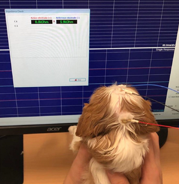 In
a
January 2023 publication of a Brazilian veterinary doctoral thesis
by Tatiana Asunción de Moraes, she reports studying 50 cavalier King
Charles spaniel puppies between the ages of 20 and 90 days plus ages 1
and 2 years, to determine at what age the CKCS achieves visual
maturation. She used Sweep-Visual Evoked Potential (S-VEP), an
electrophysiological tool which expresses the electrical activity of the
visual pathways up to the optic nerve to the calcarine cortex. They are
capable of detecting neuronal pool activity responding to stimuli
independently of the consciousness and attention state of the patient.
VEPs are a method to assess visual acuity (VA) in non-verbal patients
and for surgical monitoring. She divided the 50 puppies into five groups
of ten each by age -- G1 (20 days of age), G2 (30 days), G3 (45 days),
G4 (90 days), G5 (1 to 2 years old). She reported finding a statistical
difference between groups G1 x G3, and G2 x G3, unlike G3 x G4 x G5,
that did not show differences between these three older groups,
suggesting a plateau of VA values. Based on the results, she concluded
that the visual maturity of the cavaliers studied was achieved by 30 to
45 days of age.
In
a
January 2023 publication of a Brazilian veterinary doctoral thesis
by Tatiana Asunción de Moraes, she reports studying 50 cavalier King
Charles spaniel puppies between the ages of 20 and 90 days plus ages 1
and 2 years, to determine at what age the CKCS achieves visual
maturation. She used Sweep-Visual Evoked Potential (S-VEP), an
electrophysiological tool which expresses the electrical activity of the
visual pathways up to the optic nerve to the calcarine cortex. They are
capable of detecting neuronal pool activity responding to stimuli
independently of the consciousness and attention state of the patient.
VEPs are a method to assess visual acuity (VA) in non-verbal patients
and for surgical monitoring. She divided the 50 puppies into five groups
of ten each by age -- G1 (20 days of age), G2 (30 days), G3 (45 days),
G4 (90 days), G5 (1 to 2 years old). She reported finding a statistical
difference between groups G1 x G3, and G2 x G3, unlike G3 x G4 x G5,
that did not show differences between these three older groups,
suggesting a plateau of VA values. Based on the results, she concluded
that the visual maturity of the cavaliers studied was achieved by 30 to
45 days of age.
July 2022:
USA researchers find evidence dogs can 'see' with their sense of
smell.
In a
July 2022 article, researchers from Cornell Univ. veterinary school
and Johns Hopkins Univ. (Erica F. Andrews, Raluca Pascalau, Alexandra
Horowitz, Gillian M. Lawrence, Philippa J. Johnson)
examined the olfactory systems of
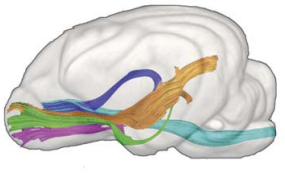 23 laboratory dogs to determine the
extent of tracts connecting the dogs; olfactory bulbs with regions of
their brains. They proceeded upon the theory of an integration of smell
and vision in dog cognition. They used diffusion tensor imaging (DTT)
MRI and a process for dissecting brain fibers, known as the Klingler
method. They report finding five tracts of "white matter" connecting the
olfactory bulb to the cortex of the brain. The tracts are: (1)
olfactory-occipital tract (OOT) (orange in diagram)
(the occipital lobe being the visual processing center of the dog's
brain); (2) olfactory-cortical spinal tract
(OCST) (light blue in diagram); (3) olfactory-limbic tract (OLT)
(dark blue in diagram); (4) olfactory-piriform tract
(OPT) (green in diagram); and (5) olfactory-entorhinal tract (OET)
(purple in diagram). They concluded
that:
23 laboratory dogs to determine the
extent of tracts connecting the dogs; olfactory bulbs with regions of
their brains. They proceeded upon the theory of an integration of smell
and vision in dog cognition. They used diffusion tensor imaging (DTT)
MRI and a process for dissecting brain fibers, known as the Klingler
method. They report finding five tracts of "white matter" connecting the
olfactory bulb to the cortex of the brain. The tracts are: (1)
olfactory-occipital tract (OOT) (orange in diagram)
(the occipital lobe being the visual processing center of the dog's
brain); (2) olfactory-cortical spinal tract
(OCST) (light blue in diagram); (3) olfactory-limbic tract (OLT)
(dark blue in diagram); (4) olfactory-piriform tract
(OPT) (green in diagram); and (5) olfactory-entorhinal tract (OET)
(purple in diagram). They concluded
that:
"These findings show the dog's olfactory system is integrated with many different parts of the brain. The OOT [occipital cortex] connection is of particular interest due to its size and relevance to understanding canine cognition. The presence of an olfactory-occipital 'information highway' provides structural evidenc for the theorized olfactory-vision integration proposed in canine cognitive research."
Researcher Dr. Johnson (right) explained about seeing with their noses, in an interview about her article:
"We've never seen this connection between the nose and the occipital lobe, functionally the visual cortex in dogs, in any species. When we walk into a room, we primarily use our vision to work out where the door is, who's in the room, where the table is. Whereas in dogs, this study shows that olfaction is really integrated with vision in terms of how they learn about their environment and orient themselves in it. They can still play fetch and navigate their surroundings much better than humans with the same condition. Knowing there's that information freeway going between those two areas could be hugely comforting to owners of dogs with incurable eye diseases. Veterinarians have long wondered how dogs with complete blindness have navigated so well in their environment, even in foreign and new environments. The olfactory connection we identified gives us an answer to this, and shows that they are less dependent on their eyes alone and likely use olfactory information to help navigate their world." (Emphasis added.)
June 2021:
USA opthalmologists use computed tomography (CT) and a 3-D model
of a cyst in a cavalier to aid in eye cyst removal.
In
a
June 2021 article, a team of opthalmologist surgeons and others
(Jessica B. Burn, András M. Komáromy, Dodd G. Sledge, Rebecca Smedley,
Sarah E. Coe, Sun Young Kim) at Michigan State University report the
successful surgical removal of a conjunctival cyst in the eye of a
cavalier King Charles spaniel (right), using both computed
tomography (CT) and digital 3-D surface modeling of the cyst region to
plan a minimally invasive approach to remove the cyst while sparing the
eye. They conclude that, "Our surgical outcome was excellent with normal
function of the nasolacrimal duct and no postoperative complications."
Read more about conjunctival cysts at this link.
October 2019:
UK canine geneticists and ophthalmologists create a consortium
to research inherited eye diseases.
 On
October 15, 2019, the Animal Health Trust (AHT) announced the
creation of CRIEDD (Consortium to Research Inherited Eye Diseases in
Dogs), which includes AHT canine geneticists led by Cathryn Mellersh
(right), and six veterinary ophthalmologists, Sheila Crispin,
Stuart Ellis, Lorraine Fleming, David Gould, Christine Heinrich, and
James Oliver. The team intends to focus upon investigating current and
emerging inherited eye diseases and developing new DNA tests to benefit
a number of breeds, effectively reducing the number of affected and
carrier dogs. To kick off this research, AHT asks that DNA samples be
submitted ot its genetics laboratory. Samples can be submitted from any
dog of any breed displaying clinical signs consistent with any inherited
eye disease that have been confirmed by a veterinary ophthalmologist.
DNA samples can be collected using simple buccal swabs, which are
available, free of charge, from the Animal Health Trust, and can be
returned using the regular mail service. AHT's genetics team will screen
all DNA samples for around 80 different known inherited eye disease
causing mutations, regardless of breed or disease. For more information
please email the team at
criedd@aht.org.uk
On
October 15, 2019, the Animal Health Trust (AHT) announced the
creation of CRIEDD (Consortium to Research Inherited Eye Diseases in
Dogs), which includes AHT canine geneticists led by Cathryn Mellersh
(right), and six veterinary ophthalmologists, Sheila Crispin,
Stuart Ellis, Lorraine Fleming, David Gould, Christine Heinrich, and
James Oliver. The team intends to focus upon investigating current and
emerging inherited eye diseases and developing new DNA tests to benefit
a number of breeds, effectively reducing the number of affected and
carrier dogs. To kick off this research, AHT asks that DNA samples be
submitted ot its genetics laboratory. Samples can be submitted from any
dog of any breed displaying clinical signs consistent with any inherited
eye disease that have been confirmed by a veterinary ophthalmologist.
DNA samples can be collected using simple buccal swabs, which are
available, free of charge, from the Animal Health Trust, and can be
returned using the regular mail service. AHT's genetics team will screen
all DNA samples for around 80 different known inherited eye disease
causing mutations, regardless of breed or disease. For more information
please email the team at
criedd@aht.org.uk
November 2016: Cavalier eye sizes are large for small dogs but fit in the medium dog group. In a November 2016 article, UK ophthalmologists (Carolin L. H. Chiwitt, Stephen J. Baines, Paul Mahoney, Andrew Tanner, Christine L. Heinrich, Michael Rhodes, Heidi J. Featherstone) found that the size of the eyes of the cavalier King Charles spaniel, a small dog, was larger than other small dogs and fit within the eye size of medium sized dogs. The researchers measured the eye sizes of 100 dogs in ten breeds, incuding 10 cavalier King Charles spaniels.
The three different measurement planes of the eye which the
researchers used are the sagittal, the equatorial, and the horizontal,
as this figure shows:
The researchers used both B-mode ocular ultrasound and computed tomography (CT) to do so in a procedure called "ocular biometry", which is the measurement of the dimensions of the eye, its components, and their interrelationships. They divided the 100 dogs into three size groups -- small, medium, and large based upon the UK Kennel Club assignments -- and placed the CKCS in the small dog group. They measured eye length, width, and height using the both of the devices -- ultrasound and CT.
They found that the CKCS had a relatively large eye for the small breed group, and there was no significant difference between the CKCS eye and those in the medium breed group. They also found that their measurements using both B-mode ultrasound and CT can be applied for the diagnosis of microphthalmos and buphthalmos.
November 2016: Brazilian researchers use digital video imaging to determine eye blink rates in cavaliers. In an October 2016 abstract, a team of Brazilian ophthalmologists (GV Fontinhas Netto, AR Eyherabide, AP Ribeiro, AA Bolzan) used digital video imaging to determine the eye blink rates per minute (EBR) in eleven healthy cavalier King Charles spaniels. They found no significant difference between the left and right eyes, and the mean value of EBR was 9.6 blinks/minute. They also measured the central corneal sensitivity by evaluating the corneal touch threshold (CTT) with the Cochet-Bonnet aesthesiometer, and tear production using the Schirmer tear test 1 (STT1). Mean ± SD tear production was 24.1 ± 1.6 mm/minute and CTT was 1.7 ± 0.3 cm. All tests were performed without anesthetic eye drops.
RETURN TO TOP
Veterinary Resources:
Imperforate and micro-lachrymal puncta in the dog. K. C. Barnett. J. Sm. Anim. Pract. August 1979;20(8):481-490. Quote: Lachrymation, the excessive secretion of tears, and epiphora, the inadequate drainage of tears, are common presenting clinical signs. It has long been supposed that the majority of cases of epiphora occur because part, or parts, of the naso-lachrymal duct system is congenitally absent or blocked and, if the former, that treatment is not possible. Imperforate and micro-lachrymal puncta are reported in detail in the dog. The main breeds affected are the Golden Retriever and Cocker Spaniel. Diagnosis and surgical treatment are discussed. The condition is considered to be more common than is realized. ... Cases in this series occured in the yellow Labrador (three), Samoyed (two) and single cases in the American Cocker Spaniel, English Springer Spaniel, Cavalier King Charles Spaniel, ... Case 26: Cavalier King Charles -- tri --- M -- 6 mos -- microphthalmos of upper and lower right lids.
Reticulosis of the eyes and the central nervous system in a dog. NL Garmer, P Naeser, AJ Bergman. J.Small Animal Prac.; Jan. 1981;22(1):39-45. Quote: "Bilateral optic neuritis was diagnosed in a 5-year old dog, which had been blind for two days. ... CASE REPORT A 5-year old, 10 kg, male Cavalier King Charles Spaniel was examined because the dog had appeared blind for two days. ... Vision returned after corticosteroid therapy. Two weeks after the end of this treatment the dog became blind again and, in addition, showed ataxia. Post-mortem examination revealed changes in both the central nervous system and the eyes. The histopathological changes observed were consistent with those of reticulosis.
Posterior lenticonus, cataracts and microphthalmia: congenital ocular defects in the Cavalier King Charles Spaniel. Narfstrom K, Dubielzig R. J. Sm. Anim. Pract. November 1984; doi: 10.1111/j.1748-5827.1984.tb03380.x. Quote: Eleven cases of congenital ocular defects were found in the screening of 144 Cavalier King Charles Spaniels in Sweden. Mainly posterior lenticonus, cataracts and microphthalmia were observed in the affected dogs, most of which were interrelated. Pathology was obtained from one of the cases demonstrating bilateral posterior lens capsule rupture with an unusual cellular reaction of the exposed lens material. ... The lenticular abnormalities found in the Cavalier King Charles Spaniel breed of dog mainly affect the nucleus, posterior cortex and the posterior capsule. These defects, together with an intact posterior lens capsule, indicate an early developmental insult in the formation of the foetal lens. Numerous theories have been proposed to explain posterior lenticonus formation. Most of the histologic evidence supports a congenital defect involving an abnormal embryonic remnant in the epithelial cell proliferation before the lens capsule forms or a hyperplasia of the lens accompanied by a thinning of the capsule over the defect. In the Cavalier dog the latter explanation seems to be the most likely. Persistence of hyaloid vessels or remnants of these is further evidence of an early developmental aberration. Microphthalmia is a common finding in conjunction with other severe ocular malformations which were also found in the present study.
Ehlers-Danlos syndrome in a dog: ocular, cutaneous and articular abnormalities. KC Barnett, BD Cottrell. J.Small Animal Prac. - Journal of Small Animal Practice; Oct. 1987;28(10):941-946. Quote: "Ehlers-Danlos syndrome (EDS) is an hereditary connective tissue disease in man in which the skin is easily torn. A similar condition has been described in dogs and other animals. This case report records a case in the United Kingdom in which the whole syndrome was exhibited: skin fragility, joint laxity and ocular signs of bilateral lens luxation, cataract and corneal oedema. It is the first report of ocular signs in EDS in the dog and joint laxity has been reported only rarely. ... A crossbred bitch (Cavalier King Charles Spaniel x Rorder Collie bitch) was presented at the age of 12 months for investigation of failing vision. ... The case reported here was a Cavalier King Charles Spaniel x Border Collie; neither breed has previously been implicated."
Angiostrongylus vasorum in the anterior chamber of a dog's eye. M. C. A. King, R. M. R. Grose, G. Startup. J. Small Anim. Prac. June 1994;35:326-8. Quote: "An unusual case of Angiostrongylus vasorum infestation occurred in a three-year-old female cavalier King Charles spaniel. The dog presented with signs consistent with right otitis interna, followed by the appearance of a free-swimming nematode in the anterior chamber of the right eye. The dog died of acute heart failure before surgical removal of the parasite was possible. Post mortem examination confirmed the presence of large numbers of worms in the pulmonary artery and right ventricle. These worms were identified histologically as A vasorum." Age, sex and breed: 3-year-old female Cavalier King Charles Spaniel. Location: UK. Clinical presentation: Otitis interna, head tilt, submaxillary lymph node enlargement. Presence of a motile worm in the anterior chamber of the eye on a one-day follow up visit. The dog died from a sudden attack of acute respiratory distress. Diagnosis: Numerous A. vasorum adults in the right ventricle and pulmonary artery and one adult in the anterior chamber detected at post-mortem examination.
Control of Canine Genetic Diseases. Padgett, G.A., Howell Book House 1998, pp. 198-199, 239.
Ocular Disorders Presumed to be Inherited in Purebred Dogs. Genetics Committee, A.C.V.O. 1999.
Guide to Congenital and Heritable Disorders in Dogs. Dodds WJ, Hall S, Inks K, A.V.A.R., Jan 2004, Section II(88).
Optic neuritis in a Cavalier King Charles Spaniel. Zarfoss, M. K. Cornell College of Vet. Med. February 2004;Seminar SF610.1 2004 Z37. Quote: Betsy, a 4.5 year-old female spayed Cavalier King Charles Spaniel, was presented to the Cornell University Hospital for Animals for evaluation of acute blindness, inappetance, and lethargy. Ophthalmic examination revealed keratoconjunctivitis sicca in both eyes (OU), absent menace OU, decreased pupillary light responses OU, posterior vitreal haze OU, and optic disk enlargement OU. A diagnosis of optic neuropathy was pursued because of bilaterally enlarged optic disks in the absence of other localizing clinical signs or ophthalmic abnormalities sufficient to cause blindness. The presumptive diagnosis of optic neuritis due to granulomatous meningo-encephalomyelitis (GME) was based upon: peripapillary retinal vasculitis on fluorescein angiography, severe lymphocytic inflammation within the cerebrospinal fluid (CSF), negative infectious disease serum and CSF titers, and favorable response to immunosuppression. Granulomatous meningoencephalomyelitis is an inflammatory disease of the central nervous system (CNS) of undetermined etiology and poor prognosis. Successful treated of this dog's GME consisted of conventional treatment using oral prednisone and non-conventional treatment using an additional immunosuppressive agent, cytosine arabinoside.
Breed Predispositions to Disease in Dogs & Cats. Alex Gough, Alison Thomas. 2004; Blackwell Publ. 44-45.
Angiostrongylus vasorum at a pre-adult phase in the anterior chamber of a young dog's eye. D. Payne. Munich: International Veterinary Ophthalmology Meeting. 2004;pg.125. Age and breed: 8-month-old Cavalier King Charles Spaniel. Location: France. Clinical presentation: Cough, uveitis, and presence of a motile worm in the anterior chamber of the eye. Diagnosis: Faecal and broncho-alveolar washing examination negative for A. vasorum larvae. One immature A. vasorum female extracted from the eye. Anthelmintic treatment: Fenbendazole-based treatment for 2 weeks.
Incidence of Canine Glaucoma with Goniodysplasia in Japan: A Retrospective Study. Kumiko Kato, Nobuo Sasaki, Satoru Matsunaga, Ryohei Nishimura, Hiroyuki Ogawa. J. Vet. Med. Sci. August 2006;68(8):853-858. Quote: The incidence of primary and secondary glaucoma in dogs was investigated. A total of 1244 dogs received ophthalmologic examinations, including tonometry and gonioscopy. Goniophotographs were taken using a goniolens to evaluate the iridocorneal angle (ICA) as well as pectinate ligament (PL). The anterior width of the ciliary cleft and the total distance from the origin of the PL to the anterior corneal surface were measured from the goniophotographs. Glaucoma was diagnosed based on the cupping of the optic nerve head, clinical signs, ocular changes, and high IOP, and it was synchronized with gonioscopic grades to differentiate between primary and secondary glaucoma. We investigated 1244 dogs of 29 breeds, including the mixed breed; among these, glaucoma was diagnosed in 127 dogs (162 eyes) [including two cavalier King Charles spaniels]. Of 162 eyes, primary glaucoma was diagnosed in 129 eyes and secondary glaucoma in 33 eyes. Shiba Inu dogs (42 dogs, 33%) showed the highest incidence of glaucoma, followed by Shih-Tzu (21 dogs, 16.5%). Furthermore, all the glaucomatous Shiba Inu dogs had primary glaucoma with abnormal ICA grades and dysplastic PLs. The findings of our study reveal that the Shiba Inu breed in Japan may have a hereditary predisposition to glaucoma.
Corneal squamous cell carcinoma in dogs with a history of chronic keratitis. R. R. Dubielzig, C. S. Schobert and J. Dreyfus. Vet Ophth; 2008;11(6):413-429 (Abstract 101). Quote: Purpose: Corneal squamous cell carcinoma (SCC) is a rare tumor in dogs. The COPLOW has seen a recent increase in primary SCC in the axial cornea. We report here on 25 cases. Methods: Twenty-five cases of primary axial corneal SCC were selected from the COPLOW collection which includes more that 6000 neoplastic specimens. ... Results: The number of canine corneal SCC has risen in the past several years from 1 case per year from 1998 to 2004, jumping to 6 cases in 2005, 8 cases in 2006, and 7 cases in 2007. Brachycephalic breeds are overrepresented. The breed distribution included 8 Pugs, 5 Bulldog, 2 Boxers, 2 Greyhound, 2 Shi Tzu, 2 Border Collie, 2 Pekinese, 1 Bassett, 1 Chow, 1 Cocker, and 1 Cavalier King Charles Spaniel. No correlation to sex was found. Out of the 25 cases, 21 showed signs of chronic keratitis prior to developing SCC. In the remaining 4 cases the prior corneal history was unknown. Within the group of 25, 10 cases had been treated with cyclosporine alone, 4 with tacrolimus alone, 5 with both cyclosporine and tacrolimus, and 6 treated with other drugs or unknown. Follow-up information was obtained from 23 cases with a follow-up interval of between 5 days and 31 months (mean: 7.9 months). Three dogs had died for reasons unrelated to the ocular disease. One dog had recurrent disease extending deeply into the cornea. Conclusions: Brachycephalic dogs with a background of chronic keratitis that are treated with nonsteroidal antiinflammatory drugs are at risk to develop axial corneal SCC. The increase in annual cases of SCC suggests that this phenomenon is a developing problem.
Ophthalmic Disease in Veterinary Medicine. Charles L. Martin. Manson Publ. 2009; page 475, table 15.1. Quote: "Presumed Inherited Ocular Diseases: Table 15.1: Breed predisposition to eye disease in dogs: Cavalier King Charles Spaniel: ... ."
Diagnosis and Treatment of Inherited Canine Corneal Diseases. Kerry L. Ketring. NAVC Conf. June 2009. Quote: After more than 30 years in private practice, I realize the majority of canine ocular diseases have a breed predisposition. This article reflects the most common corneal diseases in the breeds seen in my practice. It is pertinent to keep in mind, however, that breed incidence may reflect just the popularity of the breed. The awareness of breed-specific corneal diseases is a head start in making the correct diagnosis. ... Corneal Dystrophy: ... Any breed may develop a corneal dystrophy but the occurrence is high in the Afghan hound, Akita, boxer, Cavalier King Charles spaniel, cocker spaniel, collie, Dalmatian, golden retriever, Labrador retriever, Lowchen, miniature pinscher, Siberian husky, and Weimaraner. ... Squamous Cell Carcinoma: pink elevated and cauliflower-like mass is seen in the axial cornea in exophthalmic breeds with a history of chronic corneal disease. The corneal disease is usually associated with KCS or exposure keratitis. This isolated mass is most commonly seen in older Cavalier King Charles spaniels, pugs, and Shih Tzus. These tumors can be removed with care by a stromal keratectomy. Laser therapy, B-radiation, and cryotherapy have been recommended following the keratectomy. I feel each is either not practical or very risky to the integrity of the cornea. Following a keratectomy, the cornea should be protected with either a third eyelid flap or temporary tarsorraphy and treated aggressively for the ulcer. Even with clean margins reported by a pathologist, I have still had recurrence. Since these are slow growing and do not tend to penetrate, benign neglect with treatment for primary disease may be adequate in some older dogs with this condition.
Breed Predispositions to Disease in Dogs & Cats (2d Ed.). Alex Gough, Alison Thomas. 2010; Blackwell Publ. 53.
Ocular conditions affecting the brachycephalic breeds. Peter G.C. Bedford. 2010. RVC. Quote: "There are two types of disease which affect the eye of the brachycephalic breeds and both are directly or indirectly related to genetic predisposition. First and by far the commonest are those conditions which are due to be conformation of skull and are related to the exophthalmos which is the common feature of these breeds. Second there are those conditions which have been unwittingly bred into some brachycephalic breeds in the pursuit of desired breed characteristics. In this lecture I will present an overview of all the diseases that the small animal practitioner is likely to encounter in the brachycephalic breeds of pedigree dog. The fourteen breeds I have included for discussion are the Affenpinscher, Boston Terrier, Boxer, Bulldog, Cavalier King Charles and the King Charles Spaniels, (mesaticephalic) French Bulldog, Griffon Bruxellois, Japanese Chin, Lhasa Apso, Pekingese, Pug, Shih Tzu and Tibetan Spaniel. ... Corneal Lipid Dystrophy: The term applies to the characteristic cholesterol and triglyceride deposits in the superficial corneal stroma seen most commonly in the Cavalier King Charles Spaniel. It is clinically benign and seldom affects vision to any noticeable degree. ... Hereditary Cataract: Hereditary cataract is seen in the Boston Terrier and the Cavalier King Charles Spaniel. ... Microphthalmos (MoD): Again the American literature suggests that microphthalmos (MoD) may be inherited in the Cavalier King Charles Spaniel."
Congenital keratoconjunctivitis sicca and ichthyosiform dermatosis in 25 Cavalier King Charles spaniel dogs - part I: clinical signs, histopathology, and inheritance. Claudia Hartley, David Donaldson, Ken C. Smith, William Henley, Tom W. Lewis, Sarah Blott, Cathryn Mellersh, Keith C. Barnett. Vet.Opht. January 2011; doi:10.1111/j.1463-5224.2011.00986.x. Quote: The clinical presentation and progression (over 9 months to 13 years) of congenital keratoconjunctivitis sicca and ichthyosiform dermatosis (CKCSID) in the Cavalier King Charles spaniel dog are described for six new cases and six previously described cases. Cases presented with a congenitally abnormal (rough/curly) coat and signs of KCS from eyelid opening. Persistent scale along the dorsal spine and flanks with a harsh frizzy and alopecic coat was evident in the first few months of life. Ventral abdominal skin was hyperpigmented and hyperkeratinized in adulthood. Footpads were hyperkeratinized from young adulthood with nail growth abnormalities and intermittent sloughing. Long-term follow-up of cases (13/25) is described. Immuno-modulatory/lacrimostimulant treatment had no statistically significant effect on Schirmer tear test results, although subjectively, this treatment reduced progression of the keratitis. Histopathological analysis of samples (skin/footpads/ lacrimal glands/salivary glands) for three new cases was consistent with an ichthyosiform dermatosis, with no pathology of the salivary or lacrimal glands identified histologically. Pedigree analysis suggests the syndrome is inherited by an autosomal recessive mode.
Epidemiology of canine glaucoma presented to University of Zurich from 1995 to 2009. Part 2: secondary glaucoma (217 cases). Ann Refstrup Strom, Michael Hassig, Tine M. Iburg, Bernhard M. Spiess. Vet. Ophth. March 2011;14(2):127-132. Quote: Objective: To investigate the epidemiology of canine secondary glaucomas in the cases presented to the University of Zurich, Vetsuisse Faculty (UZH) from 1995 to 2009 focusing on possible risk factors for developing secondary glaucoma in this population of dogs. Methods: Information was obtained from the computer database of patients examined by members of the UZH Ophthalmology Service, between January 1995 and August 2009. Secondary glaucoma was diagnosed based on the presence of antecedent eye conditions. The data was evaluated for breed, gender, age at presentation, and for antecedent eye conditions known to cause glaucoma including anterior uveitis of unknown cause (AU), lens luxation (LL), intraocular surgery (SX), intraocular neoplasia (IN), unspecified trauma to the globe (T), ocular melanosis (OM), hypermature cataract (PY), hyphema (HY), and six other less frequent conditions. Results: A total of 217 dogs were diagnosed with secondary glaucoma from 1995 to 2009. The age of the dogs with secondary glaucoma ranged between 88 days and 19 years (mean 7.7 ± 3.6 years). Data suggested a predisposition for secondary glaucoma in the Cairn Terrier and the Jack Russell Terrier breeds from 2004 to 2009. Common causes of secondary glaucoma from 1995 to 2009 were AU (23.0%), LL (22.6%), SX (13.4%), IN (10.6%), T (8.3%), OM and PY (both 6.9%) and HY (3.23%). Conclusion: The report presents the epidemiology of secondary glaucomas presented to UZH from 1995 to 2009. ... The proportion of the Cairn Terriers, Jack Russell Terriers, Cocker Spaniels, Cavalier King Charles Spaniels, and Manchester Terriers in SEC0409 [secondary glaucoma group] were all overrepresented compared with the proportion of the same breeds in GAO409 [all the dogs presented to the hospital]. [Of 72 CKCSs presented to the hospital, 2 were diagnosed with secondary glaucoma.] ... Fourteen risk factors were recorded for secondary glaucoma. This is the first paper documenting OM in the Swiss Cairn Terrier dog population.
Superficial corneal squamous cell carcinoma occurring in dogs with chronic keratitis. Jennifer Dreyfus, Charles S. Schobert, Richard R. Dubielzig. Vet Ophth; May 2011;14(3):161-168. Quote: Objective: Canine corneal squamous cell carcinoma (SCC) is a rare tumor, with only eight cases previously published in the veterinary literature. The Comparative Ocular Pathology Lab of Wisconsin (COPLOW) has diagnosed 26 spontaneously occurring cases, 23 in the past 4 years [three of which were cavalier King Charles spaniels]. This retrospective study describes age and breed prevalence, concurrent therapy, biologic behavior, tumor size and character, and 6-month survival rates after diagnosis. Results: A search of the COPLOW database identified 26 corneal SCC cases diagnosed from 1978 to 2008. There is a strong breed predilection (77%) in brachycephalic breeds, particularly those prone to keratoconjunctivitis sicca. The mean age was 9.6 years (range 6-14.5 years). Follow-up information >6 months was available for 15 of 26 cases. Recurrence occurred in the same eye in nine cases, seven of which were incompletely excised at the time of first keratectomy. No cases were known to have tumor growth in the contralateral eye and no cases of distant metastases are known. Where drug history is known, 16 of 21 dogs had a history of treatment with topical immunosuppressive therapy (cyclosporine or tacrolimus) at the time of diagnosis. Conclusion: Chronic inflammatory conditions of the cornea and topical immunosuppressive therapy may be risk factors for developing primary corneal SCC in dogs. SCC should be considered in any differential diagnosis of corneal proliferative lesions. Superficial keratectomy with complete excision is recommended, and the metastatic potential appears to be low.
Ocular Disorders Presumed to be inherited in purebred dogs. Genetics Committee of the American College of Veterinary Ophthalmologists. Blue Book 6th Ed. 2013. pp. 241-247.
Blindness: A Step-by-Step Approach to Diagnosis. Alison Clode. NAVC Clinician's Brief. April 2014;14-15.
The genetics of eye disorders in the dog. Cathryn S. Mellersh. Canine Genetics & Epidemiology. April 2014. Quote: "Inherited forms of eye disease are arguably the best described and best characterized of all inherited diseases in the dog, at both the clinical and molecular level and at the time of writing 29 different mutations have been documented in the scientific literature that are associated with an inherited ocular disorder in the dog. The dog has already played an important role in the identification of genes that are important for ocular development and function as well as emerging therapies for inherited blindness in humans. Similarities in disease phenotype and eye structure and function between dog and man, together with the increasingly sophisticated genetic tools that are available for the dog, mean that the dog is likely to play an ever increasing role in both our understanding of the normal functioning of the eye and in our ability to treat inherited eye disorders. This review summarises the mutations that have been associated with inherited eye disorders in the dog."
Syringomyelia: determining risk and protective factors in the conformation
of the Cavalier King Charles Spaniel dog.
Thomas
J. Mitchell, Susan P. Knowler, Henny van den Berg, Jane Sykes, Clare
Rusbridge. Canine Genetics & Epidemiology. July 2014. Quote: "Syringomyelia
(SM) is a painful condition, more common in toy breeds, including the
Cavalier King Charles Spaniel (CKCS), than other breeds. In
these toy breeds, SM is usually secondary to a specific malformation of the
skull (called Chiari-like Malformation, CM for short). There has been debate
as to whether head shape is related to CM/SM, especially as some humans have
similar characteristic facial and skull shapes, and
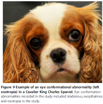 what this may be.
Identifying a head shape in dogs that is associated with these diseases
would allow for selection away from these conditions and could be used to
further breeding guidelines. 133 dogs were measured in several countries
using a standardised 'bony landmark' measuring system and photo analysis by
trained researchers. This paper describes two significant risk factors
associated with CM/SM in the skull shape of the CKCS:
extent of brachycephaly (the broadness of the cranium (top of skull)
relative to its length) and distribution of doming of the cranium. The study
showed that having a decreased cephalic index (less brachycephaly) was
significantly protective. Further to this, more cranium at the back of the
head (caudally) relative to the amount at the front of the head (rostrally)
was significantly protective against disease development. This was shown at
three and five years of age, and also when comparing a sample of "SM clear"
individuals over five years to those affected under three years. This study
suggests that brachycephaly, with resulting rostrocaudal doming, is
associated with CM/SM. ... Eye conformational abnormalities,
including strabismus, exophthalmos and exotropia determined
byobservation when dog looking forwards, left and right (Figure 9).
... These results could provide a way for selection
against the risk head shape in the CKCS, and thus enable a
reduction in CM/SM incidence. Studying other breeds in which CM free
individuals are more frequent may validate this risk phenotype for CM too."
what this may be.
Identifying a head shape in dogs that is associated with these diseases
would allow for selection away from these conditions and could be used to
further breeding guidelines. 133 dogs were measured in several countries
using a standardised 'bony landmark' measuring system and photo analysis by
trained researchers. This paper describes two significant risk factors
associated with CM/SM in the skull shape of the CKCS:
extent of brachycephaly (the broadness of the cranium (top of skull)
relative to its length) and distribution of doming of the cranium. The study
showed that having a decreased cephalic index (less brachycephaly) was
significantly protective. Further to this, more cranium at the back of the
head (caudally) relative to the amount at the front of the head (rostrally)
was significantly protective against disease development. This was shown at
three and five years of age, and also when comparing a sample of "SM clear"
individuals over five years to those affected under three years. This study
suggests that brachycephaly, with resulting rostrocaudal doming, is
associated with CM/SM. ... Eye conformational abnormalities,
including strabismus, exophthalmos and exotropia determined
byobservation when dog looking forwards, left and right (Figure 9).
... These results could provide a way for selection
against the risk head shape in the CKCS, and thus enable a
reduction in CM/SM incidence. Studying other breeds in which CM free
individuals are more frequent may validate this risk phenotype for CM too."
Impact of Facial Conformation on Canine Health: Corneal
Ulceration. Rowena M. A. Packer, Anke Hendricks,
Charlotte C. Burn. PlosOne. May 2015. Quote: Concern has arisen
in recent years that selection for extreme facial morphology in
the domestic dog may be leading to an increased frequency of eye
disorders. Corneal ulcers are a common and painful eye problem
in domestic dogs that can lead to scarring and/or perforation of
the cornea, potentially causing blindness. Exaggerated
juvenile-like craniofacial conformations and wide eyes have been
suspected as risk factors for corneal ulceration. ... Several
brachycephalic breeds have been identified as being predisposed
to dry eye, including the Bulldog, Lhasa Apso, Shih Tzu, Pug,
Pekingese, Boston Terrier and Cavalier King Charles
Spaniel. Even moderately lowered tear production
associated with dry eye may produce clinical signs in
brachycephalic dogs, as a larger portion of the globe is
exposed. In a UK based study, a higher proportion of
brachycephalic dogs that were affected by ulcers, than were
non-brachycephalic dogs with dry eye, e.g. 36% of Shih Tzus and
30% of Cavalier King Charles Spaniels versus
17% of dogs in the overall study population. ... This study
aimed to quantify the relationship between corneal ulceration
risk and conformational factors including relative eyelid
aperture width, brachycephalic (short-muzzled) skull shape, the
presence of a nasal fold (wrinkle), and exposed eye-white. A 14
month cross-sectional study of dogs entering a large UK based
small animal referral hospital for both corneal ulcers and
unrelated disorders was carried out. Dogs were classed as
affected if they were diagnosed with a corneal ulcer using
fluorescein dye while at the hospital (whether referred for this
disorder or not), or if a previous diagnosis of corneal ulcer(s)
was documented in the dogs' histories. Of 700 dogs recruited, measured and clinically examined, 31 were affected by corneal
ulcers. Most cases were male (71%), small breed dogs (mean± SE
weight: 11.4±1.1 kg), the most common being the Pug (n = 12
affected), the Shih Tzu (n = 4), the Bulldog and the
Cavalier King Charles Spaniel (n = 3). ... Morphometric
data were collected for each dog using previously defined
measuring protocols, measuring 11 conformational features
that were demonstrated to be breed-defining: muzzle length,
cranial length, head width, eye width, neck length, neck girth,
chest girth, chest width, body length, height at the withers and
height at the base of tail (all in cm). ... A further
morphometric predictor of interest for corneal ulcers was
craniofacial ratio, (CFR): the muzzle length divided by the
cranial length, which quantifies the degree of brachycephaly,
was used to differentiate skull morphologies. [Photos at right:
"This Cavalier King Charles
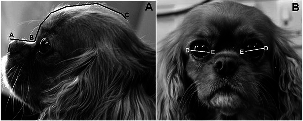 Spaniel
has a craniofacial ratio of 0.27 (muzzle length 28mm /
cranial length 102mm), and a relative palpebral fissure width
value of 33.3% ((palpebral fissure width 34mm / cranial length
102mm) *100"] ... [B]rachycephalic dogs (craniofacial
ratio <0.5) were twenty times more likely to be affected than
non-brachycephalic dogs. A 10% increase in relative eyelid
aperture width more than tripled the ulcer risk. Exposed
eye-white was associated with a nearly three times increased
risk. The results demonstrate that artificially selecting for
these facial characteristics greatly heightens the risk of
corneal ulcers, and such selection should thus be discouraged to
improve canine welfare.
Spaniel
has a craniofacial ratio of 0.27 (muzzle length 28mm /
cranial length 102mm), and a relative palpebral fissure width
value of 33.3% ((palpebral fissure width 34mm / cranial length
102mm) *100"] ... [B]rachycephalic dogs (craniofacial
ratio <0.5) were twenty times more likely to be affected than
non-brachycephalic dogs. A 10% increase in relative eyelid
aperture width more than tripled the ulcer risk. Exposed
eye-white was associated with a nearly three times increased
risk. The results demonstrate that artificially selecting for
these facial characteristics greatly heightens the risk of
corneal ulcers, and such selection should thus be discouraged to
improve canine welfare.
Keratomycosis in five dogs. Jessica C. Nevile, Simon D. Hurn, Andrew G. Turner. Vet. Ophthalmology. September 2015. Quote: "Five cases of canine keratomycosis were diagnosed and treated at a private Veterinary Ophthalmology Practice in Melbourne, Australia. Clinical presentations varied between dogs. Predisposing factors were identified in 4 of 5 cases. Diagnostic modalities utilized were corneal cytology and fungal culture. ... Case 1. A 10-year 7-month-old spayed female Cavalier King Charles Spaniel was referred for a corneal ulcer in the right eye (OD). The dog was systemically healthy, there was no known history of trauma, and the dog had not been treated with topical or systemic corticosteroids prior to presentation. ... Corneal cytology confirmed the presence of fungal organisms in all five cases. Aspergillus, Scedosporium, and Candida were cultured from three cases, respectively. Specific antifungal treatment included 1% voriconazole solution or 1% itraconazole ointment. Keratectomy and conjunctival grafting surgery was performed in two patients. Resolution of infection and preservation of vision were achieved in 4 of 5 patients."
Angiostrongylus vasorum in the eye: new case reports and a review of
the literature. Vito Colella, Riccardo Paolo Lia, Johana
Premont, Paul Gilmore, Mario Cervone, Maria Stefania Latrofa, Nunzio
D'Anna, Diana Williams, Domenico Otranto. BioMed Central. March
2016. Quote: "Background: Nematodes of the genus Angiostrongylus are
important causes of potentially life-threatening diseases in several
animal species and humans. Angiostrongylus vasorum affects the right
ventricle of the heart and the pulmonary arteries in dogs, red foxes
and other carnivores. The diagnosis of canine angiostrongylosis may
be challenging due to the wide spectrum of clinical signs. Ocular
manifestations have been seldom reported but have serious
implications for patients. Methods: The clinical history of three
cases of infection with A. vasorum in dogs diagnosed in UK, France
and Italy, was obtained from clinical records provided by the
veterinary surgeons along with information on the diagnostic
procedures and treatment. Nematodes collected from the eyes of
infected dogs were morphologically identified to the species level
and molecularly analysed by the amplification of the nuclear 18S
rRNA gene. Results: On admission, the dogs were presented with
various degrees of ocular discomfort and hyphema because of the
presence of a motile object in the eye. The three patients had
ocular surgery during which nematodes were removed and subsequently
morphologically and molecularly identified as two adult males and
one female of A. vasorum. ...
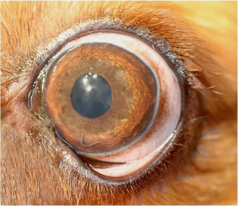 Case
2: A 2-year-old male Cavalier King Charles Spaniel
dog was referred to a private veterinary clinic in Paris (France)
due to persistent blepharospasm and epiphora. The dog that lived in
the city centre was walked in a forest nearby. On clinical
ophthalmological examination the dog showed prolapse of the
nictitating membrane, photophobia on the left eye, iris hyperaemia
and corneal oedema. The intraocular pressure was 14 mmHg in the left
eye and 16 mmHg in the right eye, the fluorescein test was negative
and the Schirmer test showed a slightly increased lacrymation. A
thread-like organism was noticed in the anterior chamber of the left
eye (photo at right), which was very motile under light
stimuli. ... Removal of the parasite was performed by anterior
chamber paracentesis and the nematode was morphologically and
molecularly processed. The specimen was identified as an
unfertilised A. vasorum female. Briefly, the female nematode
measured 16 mm in length and 0.2 mm in width; genital ducts were
coiled around the reddish intestine, which appeared visible
throughout the cuticle. However, the nematode was damaged and
further morphological features were not evaluated, with the
exception of the uteri, which lacked first-stage larvae, and the
vulva. Therefore, the dog was subjected to coprological examination
for the detection of L1 using the Baermann technique. The 18S rDNA
sequences obtained from both L1 collected from the faeces and the
female nematode displayed 100 % identity to the nucleotide sequence
of A. vasorum [GenBank: AJ920365]. The dog lacked any signs of
respiratory infection, both previously and during the observation
period. The animal was treated with fenbendazol 25 mg/Kg per os SID
for 3 weeks associated with prednisolone 0.2 mg/Kg. ... Conclusions:
Three new cases of canine ocular angiostrongylosis are reported
along with a review of other published clinical cases to improve the
diagnosis and provide clinical recommendation for this parasitic
condition. In addition, the significance of migratory patterns of
larvae inside the host body is discussed. Veterinary healthcare
workers should include canine angiostrongylosis in the differential
diagnosis of ocular diseases. ... Interestingly, all dogs suffering
from canine ocular angiostrongylosis were under the 3 years of age
(i.e. 5 months to 3 years), and of the few reports now available in
literature, three and in Case 2, involved Cavalier King
Charles Spaniel dogs. However, additional epidemiological
data are needed for a clear assessment of risk factors (e.g. breed
and age) related to the occurrence of canine ocular
angiostrongylosis."
Case
2: A 2-year-old male Cavalier King Charles Spaniel
dog was referred to a private veterinary clinic in Paris (France)
due to persistent blepharospasm and epiphora. The dog that lived in
the city centre was walked in a forest nearby. On clinical
ophthalmological examination the dog showed prolapse of the
nictitating membrane, photophobia on the left eye, iris hyperaemia
and corneal oedema. The intraocular pressure was 14 mmHg in the left
eye and 16 mmHg in the right eye, the fluorescein test was negative
and the Schirmer test showed a slightly increased lacrymation. A
thread-like organism was noticed in the anterior chamber of the left
eye (photo at right), which was very motile under light
stimuli. ... Removal of the parasite was performed by anterior
chamber paracentesis and the nematode was morphologically and
molecularly processed. The specimen was identified as an
unfertilised A. vasorum female. Briefly, the female nematode
measured 16 mm in length and 0.2 mm in width; genital ducts were
coiled around the reddish intestine, which appeared visible
throughout the cuticle. However, the nematode was damaged and
further morphological features were not evaluated, with the
exception of the uteri, which lacked first-stage larvae, and the
vulva. Therefore, the dog was subjected to coprological examination
for the detection of L1 using the Baermann technique. The 18S rDNA
sequences obtained from both L1 collected from the faeces and the
female nematode displayed 100 % identity to the nucleotide sequence
of A. vasorum [GenBank: AJ920365]. The dog lacked any signs of
respiratory infection, both previously and during the observation
period. The animal was treated with fenbendazol 25 mg/Kg per os SID
for 3 weeks associated with prednisolone 0.2 mg/Kg. ... Conclusions:
Three new cases of canine ocular angiostrongylosis are reported
along with a review of other published clinical cases to improve the
diagnosis and provide clinical recommendation for this parasitic
condition. In addition, the significance of migratory patterns of
larvae inside the host body is discussed. Veterinary healthcare
workers should include canine angiostrongylosis in the differential
diagnosis of ocular diseases. ... Interestingly, all dogs suffering
from canine ocular angiostrongylosis were under the 3 years of age
(i.e. 5 months to 3 years), and of the few reports now available in
literature, three and in Case 2, involved Cavalier King
Charles Spaniel dogs. However, additional epidemiological
data are needed for a clear assessment of risk factors (e.g. breed
and age) related to the occurrence of canine ocular
angiostrongylosis."
Infectious crystalline keratopathy in dogs and cats: clinical, in vivo confocal microscopic, histopathologic, and microbiologic features of eight cases. Eric C. Ledbetter, Patrick L. McDonough, Kay Kim. Veterinary Ophthalmology. June 2016. Quote: Objective: To describe clinical, in vivo confocal microscopic, histopathologic, and microbiologic features of canine and feline cases of infectious crystalline keratopathy (ICK). Animals studied: Six dogs [including one cavalier King Charles spaniel] and two cats with naturally acquired ICK. Procedures: Medical records of dogs and cats with a clinical diagnosis of ICK were reviewed. Signalment, medical history, clinical findings, and diagnostic evaluations were retrieved, including corneal cytology, histopathology, in vivo confocal microscopy, and microbiology results. Results: All animals presented with fine, needle-like, and branching white crystalline anterior stromal opacities emanating from corneal facets or corneal epithelial defects. Mild conjunctival hyperemia and anterior uveitis were frequently present. Concurrent ocular and systemic diseases were common, including keratoconjunctivitis sicca, corneal sequestrum, diabetes mellitus, hyperadrenocorticism, and malignant neoplasia. Bacteria, with minimal or absent leukocytes, were identified by cytology and histopathology. Histopathologically, the crystalline corneal opacities corresponded with dense accumulations of bacteria present in the interlamellar stromal spaces and forming cord-like projections within the stroma. In vivo confocal microscopy demonstrated deposits of reflective crystalline or amorphous structures within the stroma with a paucity of associated inflammatory changes. The most frequently cultured bacteria were alpha-hemolytic Streptococcus and Staphylococcus species. Resolution of clinical lesions was achieved in most cases with long-term medical or surgical therapy; however, the initiation of medical treatment was associated with an acute, dramatic onset of severe keratitis and anterior uveitis in some animals. Conclusions: Infectious crystalline keratopathy in dogs and cats shares many features with this condition in human patients. Prolonged medical therapy, or surgical intervention, is required for resolution.
Eye blink rate, tear production, and corneal sensitivity in cavalier king charles spaniels. GV Fontinhas Netto, AR Eyherabide, AP Ribeiro, AA Bolzan. Vet. Ophthal. November 2016;19(6):E25#29. Quote: Purpose: To determine the eye blink rate, tear production and corneal sensitivity in the Cavalier King Charles Spaniels. Methods: Eleven healthy Cavalier King Charles Spaniels (5 females and 6 males) aged from 3 to 10 years were included in this study. These dogs were from the "Lilies Cavaliers Kennel" (Valinhos, Sao Paulo, Brazil) and all procedures were conducted with the consent of the owner. The eye blink rate (EBR) was obtained from digital video imaging of each dog's eyes captured during three minutes and represented a counting of the eyelid movements (complete and incomplete blinks). Tear production was measured with the Schirmer tear test 1 (STT1). The central corneal sensitivity was determined by evaluating the corneal touch threshold (CTT) with the Cochet-Bonnet aesthesiometer. The tests were performed in both eyes without instillation of anesthetic eye drops. The right eye was the first eye tested. Results: There were no significant differences between the left and right eyes (P ≤ 0.05). The mean value of the EBR was 9.6 blinks/minute (0.6 complete blinks and 9.0 incomplete blinks). Mean ± SD tear production was 24.1 ± 1.6 mm/minute and CTT was 1.7 ± 0.3 cm. Conclusions: The procedures were easy to perform and reliable to measure the chosen parameters. The values obtained in our study are original (except tear production) and may contribute to establish normal patterns for eye blink rate, tear production and corneal sensitivity in adult Cavalier King Charles Spaniels.
Ocular biometry by computed tomography in different dog breeds.
Carolin L. H. Chiwitt, Stephen J. Baines, Paul Mahoney, Andrew
Tanner, Christine L. Heinrich, Michael Rhodes, Heidi J.
Featherstone. Vet. Ophth. November 2016. Quote: Objective: To
(i) correlate B-mode ocular ultrasound (US) and computed
tomography (CT) (prospective pilot study), (ii) establish a
reliable method to measure the normal canine eye using CT, (iii)
establish a reference guide for some dog breeds, (iv) compare
eye size between different breeds and breed groups, and (v)
investigate the correlation between eye dimensions and body
weight, gender, and skull type (retrospective study). Procedure:
B-mode US and CT were performed on ten sheep cadaveric eyes. CT
biometry involved 100 adult pure-bred dogs with nonocular and
nonorbital disease, representing eleven breeds [including ten
cavalier King Charles spaniels]. Eye length,
width, and height were each measured in two of three planes
(horizontal, sagittal, and equatorial). Results: B-mode US and
CT measurements of sheep cadaveric eyes correlated well
(0.70-0.71). The shape of the canine eye was found to be akin to
an oblate spheroid (a flattened sphere). A reference guide was
established for eleven breeds. Eyes of large breed dogs (GSD,
Labrador Retriever, Boxer) were significantly larger than those
of medium ((Border Collie, SBT, Cocker Spaniel, ESS) and small
breed dogs (CKCS, Border Terrier, JRT, WHWT) (P
< 0.01), and eyes of medium breed dogs were significantly larger
than those of small breed dogs (P < 0.01). ... There was no
significant difference between the CKCS eye (a
small breed) and the medium breed group. ... Eye size correlated
with body weight (0.74-0.82) but not gender or skull type.
Conclusions: Computed tomography is a suitable method for
biometry of the canine eye, and a reference guide was
established for eleven breeds. Eye size correlated with breed
size and body weight. ... The CKCS had a relatively large eye
for the small breed group. ... Because correlation between
B-mode US and CT was shown, the obtained values can be applied
in the clinical setting, for example, for the diagnosis of
microphthalmos and buphthalmos.
Use of a 350-mm2 Baerveldt glaucoma drainage device to maintain vision and control intraocular pressure in dogs with glaucoma: a retrospective study (2013-2016). Kathleen L. Graham, David Donaldson, Francis A. Billson, F. Mark Billson. Vet. Ophth. November 2016. Quote: Objective: To evaluate the 350-mm2 Baerveldt glaucoma drainage device (GDD) in dogs with refractory glaucoma when modifications to address postoperative hypotony (extraluminal ligature; intraluminal stent) and the fibroproliferative response (intraoperative Mitomycin-C; postoperative oral colchicine and prednisolone) are implemented as reported in human ophthalmology. Design: Retrospective case series. Animals: Twenty-eight client-owned dogs [including a cavalier King Charles spaniel] (32 eyes) including seven dogs (nine eyes) with primary glaucoma and 21 dogs (23 eyes) with secondary glaucoma. Methods: The medical records of all dogs undergoing placement of a 350-mm2 Baerveldt GDD at a veterinary ophthalmology referral service between 2013 and 2016 were reviewed. Signalment, diagnosis, duration and previous treatment of glaucoma, previous intraocular surgery, IOP, visual, and surgical outcomes were recorded. Results: IOP was maintained <20mmHg in 24 of 32 (75.0%) eyes. Fourteen eyes (43.8%) required no adjunctive treatments to maintain this IOP control. Fewer doses of glaucoma medication were required following surgery. Vision was retained in 18 of 27 (66.7%) eyes with vision at the time of surgery. No eyes that were blind at the time of surgery (n = 5) had restoration of functional vision. Complications following surgery included hypotony (26/32; 81.3%), intraocular hypertension (24/32; 75.0%), and fibrin formation within the anterior chamber (20/32; 62.5%). The average follow-up after placement of the GDD was 361.1 days (median 395.6 days). Conclusion: Efforts to minimize postoperative hypotony and address the fibroproliferative response following placement of a 350-mm2 Baerveldt GDD showed an increased success rate to other reports of this device in dogs and offers an alternative surgical treatment for controlling intraocular pressure in dogs with glaucoma.
Hyperfibrinolysis and Hypofibrinogenemia Diagnosed With Rotational Thromboelastometry in Dogs Naturally Infected With Angiostrongylus vasorum. N.E. Sigrist, N. Hofer-Inteeworn, R. Jud Schefer, C. Kuemmerle-Fraune, M. Schnyder, A.P.N. Kutter. J. Vet. Intern. Med. May 2017. Quote: Background: The pathomechanism of Angiostrongylus vasorum infection-associated bleeding diathesis in dogs is not fully understood. Objective: To describe rotational thromboelastometry (ROTEM) parameters in dogs naturally infected with A. vasorum and to compare ROTEM parameters between infected dogs with and without clinical signs of bleeding. Animals: A total of 21 dogs presented between 2013 and 2016. ... 3 dogs were Cavalier King Charles Spaniels (3/21, 14.3%), 2 were Chihuahuas (2/21, 9.5%), and the remaining 16 dogs represented different breeds. ... Methods: Dogs with A. vasorum infection and ROTEM evaluation were retrospectively identified. Thrombocyte counts, ROTEM parameters, clinical signs of bleeding, therapy, and survival to discharge were retrospectively retrieved from patient records and compared between dogs with and without clinical signs of bleeding. Results: Evaluation by ROTEM showed hyperfibrinolysis in 8 of 12 (67%; 95% CI, 40-86%) dogs with and 1 of 9 (11%; 95% CI, 2-44%) dogs without clinical signs of bleeding (P = .016). Hyperfibrinolysis was associated with severe hypofibrinogenemia in 6 of 10 (60%; 95% CI, 31-83%) of the cases. Hyperfibrinolysis decreased or resolved after treatment with 10-80 mg/kg tranexamic acid. Fresh frozen plasma (range, 14-60 mL/kg) normalized follow-up fibrinogen function ROTEM (FIBTEM) maximal clot firmness in 6 of 8 dogs (75%; 95% CI, 41-93%). Survival to discharge was 67% (14/21 dogs; 95% CI, 46-83%) and was not different between dogs with and without clinical signs of bleeding (P = .379). Conclusion and Clinical Importance: Hyperfibrinolysis and hypofibrinogenemia were identified as an important pathomechanism in angiostrongylosis-associated bleeding in dogs. Hyperfibrinolysis and hypofibrinogenemia were normalized by treatment with tranexamic acid and plasma transfusions, respectively.
Angiostrongylus vasorum infection in dogs from a cardiopulmonary dirofilariosis endemic area of Northwestern Italy: a case study and a retrospective data analysis. Olivieri E, Zanzani SA, Gazzonis AL, Giudice C, Brambilla P, Alberti I, Romussi S, Lombardo R, Mortellaro CM, Banco B, Vanzulli FM, Veronesi F, Manfredi MT. BMC Vet. Res. June 2017;12:165. Quote: Background: In Italy, Angiostrongylus vasorum, an emergent parasite, is being diagnosed in dogs from areas considered free of infection so far. As clinical signs are multiple and common to other diseases, its diagnosis can be challenging. In particular, in areas where angiostrongylosis and dirofilariosis overlap, a misleading diagnosis of cardiopulmonary dirofilariosis might occur even on the basis of possible misleading outcomes from diagnostic kits. Case Presentation: Two Cavalier King Charles spaniel dogs from an Italian breeding in the Northwest were referred to a private veterinary hospital with respiratory signs. A cardiopulmonary dirofilariosis was diagnosed and the dogs treated with ivermectin, but one of them died. At necropsy, pulmonary oedema, enlargement of tracheo-bronchial lymphnodes and of cardiac right side were detected. Within the right ventricle lumen, adults of A. vasorum were found. All dogs from the same kennel were subjected to faecal examination by FLOTAC and Baermann's techniques to detect A. vasorum first stage larvae; blood analysis by Knott's for Dirofilaria immitis microfilariae, and antigenic tests for both A. vasorum (Angio Detect™) and D.immitis (DiroCHEK® Heartworm, Witness®Dirofilaria). The surviving dog with respiratory signs resulted positive for A. vasorum both at serum antigens and larval detection. Its Witness® test was low positive similarly to other four dogs from the same kennel, but false positive results due to cross reactions with A. vasorum were also considered. No dogs were found infected by A. vasorum. Eventually, the investigation was deepened by browsing the pathological database of Veterinary Pathology Laboratories at Veterinary School of Milan University through 1998-2016, where 11 cases of angiostrongylosis were described. Two out of 11 dogs had a mixed infection with Crenosoma vulpis. Conclusion: The study demonstrates the need for accurate surveys to acquire proper epidemiological data on A. vasorum infection in Northwestern Italy and for appropriate diagnostic methods. Veterinary clinicians should be warned about the occurrence of this canine parasite and the connected risk of a misleading diagnosis, particularly in areas endemic for cardiopulmonary dirofilariosis.
Anatomical features of the optic canal and the cephalic index in the cranial bone of healthy dogs. Y Ichikawa, N Kanemaki. Vet. Ophthal. October 2017. DOI: 10.1111/vop.12519; E-14. Quote: Purpose: We studied the anatomical features of the optical canal among brachycephalic, mesocephalic, and dolichocephalic dogs, which are characterized by cephalic indexes, by analyzing computed tomography (CT) images of the head in healthy dogs. Methods: Thirty-two adult healthy dogs were divided into three groups. The eight brachycephalic dogs included three Cavalier King Charles spaniels, two French bulldogs and Chihuahuas, and one Shih Tzu. The 13 mesocephalic dogs included four American cocker spaniels and Cardigan Welsh corgis, two toy poodles and Yorkshire terriers, and one Labrador retriever. The 11 dolichocephalic dogs included nine miniature dachshunds, one Great Dane and Shetland sheepdog. OsiriX Lite software (v.8.0.2, Picmeo SARL, Switzerland) was used to measure the length and diameter of the optic canal and the angle of the paired canals, and cephalic index, with the CT images of the heads. Values among the groups were analyzed using a post-hoc test. Results: Stockard's and Evans's cephalic index in the brachycephalic group were 94.3 ± 13.7 and 79.4 ± 12.0, respectively, and significantly higher than those of the mesocephalic and dolichocephalic groups. The angle of paired canals in the brachycephalic, mesocephalic, and dolichocephalic groups was 103.1 ± 12.8, 82.9 ± 8.1, and 79.7 ± 5.7 degrees, respectively. There was a positive correlation between the angle of optic canals and the cephalic index. There was no significant difference in the length and diameter of the optic canal among the groups. Conclusions: The positioning of the optic canal varies with cranial morphology in dogs.
Optic Neuritis. Anansa Persaud, Thomas Chen. Clinicians Brief. March 2018;35-38. Quote: Optic neuritis is a rare but serious condition that can result in acute blindness or visual deficits in one or both eyes. Prompt diagnosis and treatment are necessary to recover vision, and evaluation for neurologic, infectious, and/or neoplastic disease may also be warranted.
Angiostrongylose oculaire chez un cavalier king charles. Mario Cervone, Laurent Bouhanna. Le Point veterinaire (ed. Expert canin). June 2018;49(384):76-79. Quote: A castrated male Cavalier King Charles spaniel was presented for exploration of blepharospasm and severe tear production in recent days. Slit lamp biomicroscopic examination revealed a living parasite in the anterior chamber of the left eye. Surgical extraction of the parasite was performed. An adult female Angiostrongylus vasorum was identified on morphological and molecular examinations. A stool examination by the Baermann method was performed to search for a pulmonary vascular parasite focus. It was positive for the presence of L1 larvae of A. vasorum. Oral fenbendazole treatment was initiated for 21 days. The ocular signs had disappeared at the 7-day postoperative check-up and the stool examination by the Baermann method proved negative for the presence of A. vasorum larvae 1 month later.
Phenobarbital-responsive bilateral zygomatic sialadenitis following an enterotomy in a cavalier king charles spaniel. Paoul S. Martinez, Renee Carter, Lorrie Gaschen, Kirk Ryan. Res. J. Vet. Practitioners. August 2018;6(2):14-19. Quote: A 6 year old male castrated Cavalier King Charles Spaniel presented to the Louisiana State University Veterinary Teaching Hospital for a three day history of bilateral acute trismus, exophthalmos, and blindness. These signs developed after an exploratory laparotomy and gastrointestinal foreign body removal was performed. On presentation, the ophthalmic examination revealed bilateral decreased retropulsion, absent bilateral direct and consensual pupillary light responses, bilateral blindness, and a superficial ulcer in right eye. Ocular ultrasound and computed tomography of the skull were performed and identified a bilateral retrobulbar mass effect originating from the zygomatic salivary glands. Ultrasound guided fine needle aspirate cytology revealed neutrophilic inflammation. Fungal testing and bacterial cultures were performed which were negative for identification of an infectious organism. The patient was placed on a 5-day course of antibiotic therapy and the response was poor. Phenobarbital was added to the treatment regimen. Two days following phenobarbital administration, the exophthalmos and trismus began to resolve. Following phenobarbital therapy, ocular position normalized, trismus completely resolved, and patient returned to normal behavior although blindness remained in both eyes. No other neurologic condition occurred. This paper highlights the early use of phenobarbital for the rapid resolution of zygomatic sialadenitis. [Zygomatic sialadenitis is an uncommon disease in dogs. It is defined as inflammation of the zygomatic salivary gland, which causes enlargement and often formation of a salivary cyst, called sialocele formation. The zygomatic gland is a salivary gland located between the nasal passage and the eyes.]
Visual Acuity Outcomes in Cavalier King Charles Spaniel Dogs by Sweep Visual Evoked Potential. TA Moraes, LMCR Carvalho, CR Paes, R Squarzoni,PY Sacai, D Otsuki, AMV Safatle. Vet. Ophthal. November 2019;22:E79-%80. Quote: Purpose: Visual acuity measurement is commonly performed in humans. In non-verbal patients, Sweep Visual Evoked Potential (s-VEP) is widely used to assessment grating visual acuity (GVA). In veterinary medicine neither reference values by age nor the moment of visual maturity was established. The aim of this study is to compare grating visual acuity by S-VEP in Cavaliers in two different ages. Methods: Ten Cavalier King Charles Spaniel dogs underwent binocular grating visual acuity measurement by s-VEP. The dogs were divided into 2 groups according to age: 30 days of life (3 females and 2 males) and one year old (5 females). All dogs had good transparency of ocular surface and normal fundoscopy. The electroencephalogram (EEG) was recorded from one from an active electrode (Oz), with the reference (Fpz) and a ground electrode (Cz). The dogs were positioned in front of the monitor, using mild manual restraint, without sedation. Results: Mean GVA upper spatial frequency limit ranged from 1.73 ± 0.09 (20/347 Snellen equivalent) in the 30 days group to 3.86 ± 0.38 (20/156) at 1 year old group. There was statistically significant difference between the groups (P < .0001). Conclusions: This study demonstrated that s-VEP is a useful tool to assess visual acuity in Cavalier King Charles Spaniel dogs. Visual acuity was better in the one year old group, suggesting the maturation process of visual pathway. More adults and puppies in different ages should be tested to extend these findings and confirm the exact moment of grating visual acuity maturation.
Diagnostic Ophthalmology. Bianca S. Bauer, Marina L. Leis, Lynne S. Sandmeyer. Can. Vet. J. March 2020;61(3):321-322. Quote: Ectopic cilia develop from undifferentiated gland tissue due to dysplasia of the meibomian (i.e., tarsal) glands. They usually arise singly or with 2 or more hairs from the meibomian glands, although occasionally a nest of cilia may be present. The follicle itself is located 4 to 6 mm behind the lid margin of the lid near the base of the meibomian gland. Unlike distichia, which arise through the meibomian duct openings at the free lid margin, ectopic cilia emerge through the palpebral conjunctiva approximately 4 to 5 mm from the lid free margin and rub directly on the cornea, causing severe corneal irritation with or without ulceration. The ectopic hairs are usually the same color as the rest of the hairs of the dog and are located in a small, pigmented spot of palpebral conjunctiva. Breeds predisposed to ectopic cilia are flat-coated retriever, Pekingese, Shih Tzu, Cavalier King Charles spaniel, boxer, English bulldog, poodle, and Jack Russell terrier. Both lids can be affected but the upper eyelid is primarily involved. The condition is usually in the young dog, accompanied by acute, intense blepharospasm, and lacrimation; similar clinical signs to a conjunctival or corneal foreign body. The hair can easily result in a superficial corneal ulcer which, with chronicity, can be accompanied by corneal blood vessels.
Presumed optic neuritis of non-infectious origin in dogs treated with immunosuppressive medication: 28 dogs (2000-2015). L. Bedos, R. Tetas, V. Crespo, A. Shea. J. Sm. Anim. Pract. September 2020; doi: 10.1111/jsap.13233. Quote: Objective: To describe the clinical findings, magnetic resonance imaging features, management and outcome of canine cases with presumed optic neuritis of non-infectious origin that were presented to a UK referral centre from January 2000 to December 2015. Materials and Methods: The clinical database was searched for optic neuritis. Dogs with acute-onset vision impairment, systemic immunosuppressive treatment and follow-up of >6 months were included. Information collected included: age; gender; breed; clinical signs and duration; physical, ophthalmic and neurological examination findings; concurrent systemic disease; and results of electroretinogram, magnetic resonance imaging, cerebrospinal fluid analysis, polymerase chain reaction and serology testing for Toxoplasma gondii, Neospora caninum and canine distemper virus, haematology and serum biochemistry profiles, abdominal ultrasound, thoracic radiography, treatment and outcome. Results: Twenty-eight dogs [including 2 cavalier King Charles spaniels (7%)] were included, with a total of 48 affected optic nerves. Age at presentation ranged from 6 months to 10.5 years. Fundoscopic evidence of optic nerve disease was present in 34 of 48 (71%) optic nerves. Magnetic resonance imaging revealed enlargement of 32 of 48 (67%) nerves and contrast enhancement of 28 of 48 (58%) nerves. Cerebrospinal fluid analysis performed in 25 of 28 (89%) dogs revealed pleocytosis (>5 nucleated cells/uL) in 11 of 25 (44%) and increased protein (>0.35 g/L) in 11 of 25 (44%). Immunosuppressive prednisolone was administered to all dogs. Prednisolone was used alone in 9 of 28 (32%) dogs; the remaining 19 dogs received a combination of prednisolone with cytosine arabinoside, cyclosporine and/or azathioprine. Vision was recovered in 24 eyes (50%) of 18 affected dogs. Clinical Significance: A positive response to treatment was observed in 64% of dogs with presumptively diagnosed optic neuritis treated with immunosuppressive medication.
Brachycephalic ocular syndrome in dogs. David Nutbrown-Hughes. UK-Vet Companion Anim. May 2021; doi: 10.12968/coan.2020.0056. Quote: Small brachycephalic breeds, such as the Pug and French Bulldog, among others, are currently extremely popular. The conformation of these breeds is part of their appeal to owners, although this can lead to ocular surface disease such as corneal ulceration and pigmentation. The eye problems associated with these breeds are collectively known as brachycephalic ocular syndrome. In dolicocephalic and mesocephalic dogs there is usually a close interaction between the tear film, the eyelids and the cornea. This does not seem to be the case in breeds with brachycephalic ocular syndrome, where poor skull and eyelid conformation, corneal sensation and tear films are associated with ocular problems such as corneal ulceration and pigmentation, as well as a predisposition to globe proptosis. Treatment needs to address the causes of the problems and combinations of both medical and surgical treatment are required.
Transconjunctival excision of an orbital conjunctival cyst using
computer-assisted 3-D surgical planning in a dog.
Jessica B. Burn, András M. Komáromy, Dodd G. Sledge, Rebecca
Smedley, Sarah E. Coe, Sun Young Kim. Clinical Case Rpts. June
2021; doi: 10.1002/ccr3.4345. Quote: Investigation of
exophthalmos and blood-colored discharge from the left ventral
punctum in a dog [a cavalier King Charles spaniel,
right]
was consistent with a conjunctival cyst in the orbit. 3-D prints
of the cyst and surrounding facial bones identified a successful
transconjunctival approach without an orbitotomy and patency of
the left lacrimal duct was reestablished. ... Conjunctival cysts
in dogs originate from either the conjunctival epithelium or the
epithelium of the lacrimal duct are frequently reported in the
orbit in dogs. They may be of primary or secondary origin, and
complete surgical excision is typically curative. The underlying
mechanism of cyst formation remains unclear; however, congenital
duct malformation, production of abnormal secretory material,
trauma, and/or inflammation leading to lacrimal gland
hypersecretion and subsequent injury to the walls of the ducts
are speculated to lead to passive dilation and cyst formation.
Successful medical and/or surgical treatments for conjunctival
cysts include injection of a sclerosing agent such as 1%
polidocanol, surgical marsupialization, and orbitotomy with
enucleation to access and surgically excise the cyst. Treatment
choice is dependent on the size and location of the cyst and the
involvement of adjacent anatomical structures. Surgical
approaches are often complicated by the presence of the globe
itself, the proximity of nerves and large vessels, and the need
to work in a small space surrounded by bones and ligaments. This
report details the surgical planning and transconjunctival
excision of a conjunctival cyst in a dog using a
patient-specific 3-D model. ... Computed tomography (CT) of the
head revealed a 2-cm-diameter, round, well-circumscribed,
thin-walled, fluid-filled structure ventromedial to the left
globe causing moderate exophthalmos with focal medial
displacement and lysis of the osseous orbital wall. ... A
digital 3-D model of the cystic lesion and its surrounding
structures including the globe, bony structures, and the
infraorbital artery was segmented [see images below], and a
partial orbitotomy was simulated to expose the cyst using 3-D
modeling software. The cystic lesion and a portion of skull
around the lesion, including the zygomatic, lacrimal, and
maxillary bones and their associated foramina, were separately
3-D printed. A simulated surgery using the 3-D digital model and
manipulation of the 3-D prints established that a
transconjunctival approach to the cyst was feasible without an
orbitotomy.
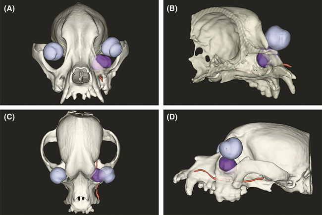
Extensive Connections of the Canine Olfactory Pathway Revealed by Tractography and Dissection. Erica F. Andrews, Raluca Pascalau, Alexandra Horowitz, Gillian M. Lawrence, Philippa J. Johnson. J. Neurosci. July 2022; doi: 10.1523/JNEUROSCI.2355-21.2022. Quote: The domestic dog's olfactory sense is widely recognized as being highly sensitive with a diverse function, however, little is known about the structure of their olfactory system. This study examined a cohort of mixed sex mesaticephalic canines and used Diffusion MRI (DTI), to map connections from the olfactory bulb to other cortical regions of the brain. The results were validated using the Klingler dissection method. ... This study identified intra-brain connections composing the extensive canine olfactory system with novel cortical connections that expands our understanding of the dominant role of olfaction has in canine congnition. ... An extensive pathway composed of five white matter tracts connecting to the occipital lobe, cortical spinal tract, limbic system, piriform lobe and entorhinal pathway was identified. This is the first documentation of a direct connection between the olfactory bulb and occipital lobe in any species and is a step towards further understanding how the dog integrates olfactory stimuli in their cognitive function. ... These findings show the dog's olfactory system is integrated with many different parts of the brain. The OOT [occipital cortex] connection is of particular interest due to its size and relevance to understanding canine cognition. The presence of an olfactory-occipital 'information highway' provides structural evidenc for the theorized olfactry-vision integration proposed in canine cognitive research. ... Significance Statement: The highly sensitive olfactory system of the domestic dog is largely unexplored. We applied diffusion tractography and dissection techniques to evaluate the white matter connections associated with the olfactory system in a large cohort of dogs. We discovered an extensive white matter network extending from the olfactory bulb to form novel connections directly to other cortices of the brain. This is the first documentation of these novel olfactory connections and provides new insight into how the dog integrates olfactory stimuli in their cognitive functioning.
Essentials of Veterinary Ophthalmology (4th ed.). Gelatt, Kirk N.; Plummer, Caryn E. Wiley-Blackwell. 2022. Quote: Dogs with distichiasis more frequently also have ectopic cilia in the dorsal conjunctiva. The cilia emerge directly on the cornea, often causing severe corneal irritation and bleparospasm. They are usually pigmented in the same color as the rest of the hairs of the dog and located in a small pigmented spot of mid-dorsal palebral conjunctiva. Pre-dispsoed breeds, also predisposed for distichiasis, are ... Cavalier King Charles Spaniel.
The pandemic of ocular surface disease in brachycephalic dogs: The brachycephalic ocular syndrome. Lionel Sebbag, Rick F. Sanchez. Vet. Ophthal. December 2022; doi: 10.1111/vop.13054. Quote: Brachycephalic dog breeds are popular around the world, yet many brachycephalic dogs are affected by numerous health problems, including several head-related diseases that are directly linked to their conformation. In addition to the well-recognized disorders associated with the respiratory system (brachycephalic obstructive airway syndrome, i.e., BOAS), brachycephalic dogs have a concerningly high prevalence of ocular surface disorders that can cause chronic discomfort, loss of the globe, and/or require long-term, daily therapy. This review offers a summary of the physiological and anatomical features of brachycephalic ocular syndrome (BOS) that predispose brachycephalic dogs to develop ocular surface disease, followed by a concise description of common ocular diseases associated with BOS. It ends with an overview of evidence-based guidelines and animal welfare legislation that some in the veterinary community have already implemented but that requires a wider, international effort in order to reduce the prevalence of BOS-associated disorders and improve the ocular health of affected dogs. ... General guidelines: Considering the evidence gathered and presented here, the following advice could be offered to dog breeders, veterinarians, and owners of brachycephalic dogs: • Breed away from extremes in facial conformation: discourage a decreased craniofacial ratio while encouraging an increased craniofacial ratio and encourage a palpebral aperture that avoids macroblepharon, a low incidence of distichiasis, ectopic cilium, and nasal fold trichiasis. • Develop and enforce breed-associated health and conservation plans for brachycephalic dogs, possibly adopting similar legislation as that developed in the Netherlands and in other countries, such as in the UK through the Kennel Club. • When facial conformation is far from ideal, and/or there are ocular complications that could be associated with brachycephaly, consider/offer owners referral for ophthalmic examination, and ultimately for anatomical correction (i.e., medial canthoplasty and nasal fold resection) and removal of distichiasis / ectopic cilium. • Offer an ophthalmic examination as part of annual examination, including STT-1, especially in breeds predisposed to DED, older patients, or dogs with ocular signs, and in dogs with mild conjunctivitis, mucoid discharge, or corneal ulceration independent of age and sex. • Consider/offer early referral in complicated cases with ocular surface disease to avoid rapidly deteriorating corneal breakdown. Until breed standards are improved over time, additional advice might be given to brachycephalic dog owners to improve their pet's ocular health, though each option would need to be further assessed in longitudinal, prospective, randomized, controlled studies. One such option would include the use topical lubrication in patients with macropalpebral fissure and lagophthalmos (i.e., a preservative-free, viscous tear supplement during the day, and the use of a topical ointment at bedtime), and regular grooming that includes periocular hair trimming to minimize the risk of inadvertent trichiasis caused by long hairs that contact the tear film. Lastly, explaining to owners in general practices and puppy classes how to train puppies to receive eye care by using gentle trimming techniques, sterile saline, or cleaning solutions that are preservative-free and have a physiologic (i.e., neutral) pH to clean debris from ocular surface and the eyelids. This would help puppies get accustomed to receiving eye care, which would improve patient and owner compliance if eye care were needed later in the dog's life.
Measurement of visual acuity of Cavalier King Charles
Spaniel dogs by evoked visual scanning potential (PVEV).
Tatiana Asunción de Moraes. Doctoral thesis, Univ. of São Paulo.
January 2023. Quote:
Visual
evaluation plays an essential role in the ophthalmic examination in
human beings. Some objective techniques are required to achieve
gratin visual acuity (VA) in non verbal people, like babies or
infants in pre-verbal ages. The Sweep-Visual Evoked Potential
(S-VEP) is suggested to be the most feasible electrophysiologic
technique to measure visual acuity. In Veterinary Medicine,
researches regarding S-VEP standadization and VA values according to
age are still scarce. The anatomical peculiarities of each breed
directly influence in visual acuity. Thus, the aim
of this study was to determine standard values of visual acuity in
Cavalier King Charles Spaniel (CKCS) dogs in
different ages and to define the moment of visual maturation in this
breed. 50 CKCS dogs were selected and divides into
five groups by age criteria: G1 (20 days of age), G2 (30 days), G3
(45 days), G4 (90 days), G5 (1 to 2 years old). The S-VEP was
performed by Roland RETIport system electrodiagnostic device and the
VA values were obtained in cicles per degree (cpd), Snellen
Equivalent, decimal and LogMAR. Statstical analysis was based on
Kruskal-Wallis test, followed by Dunn test, whenever it was
required, using the statistical software Prism 5 for Windows
(GraphPad Software). The median of the groups (from G1 to G5) were
respectly 1,250; 1,750; 3,775; 4,000 e 4,135cpg. A statistical
difference was observed between G1 x G3, and G2 x G3, unlike G3 x G4
x G5, that didn′t show differences between these three older groups,
suggesting a plateau of VA values. Based on the results, the visual
maturity of the subjects studied was achieved by 30 and 45 days of
life.
A Review of Clinical Outcomes, Owner Understanding and Satisfaction following Medial Canthoplasty in Brachycephalic Dogs in a UK Referral Setting (2016-2021). Amy L. M. M. Andrews, Katie L. Youngman, Rowena M. A. Packer, Dan G. O'Neill, Christiane Kafarnik. Animals. June 2023; doi: 10.3390/ani13122032. Quote: Brachycephalic breeds have increased in popularity despite growing awareness of their predisposition to a wide range of conformation-related diseases. The extreme facial conformation of many popular brachycephalic breeds compromises their ocular surface health, increasing the risk of painful corneal ulceration. Medial canthoplasty (MC) is a surgical procedure to address ocular abnormalities in brachycephalic dogs, which are collectively referred to as brachycephalic ocular syndrome (BOS). This study retrospectively reviewed the records of dogs recommended MC at a referral hospital between 2016 and 2021. A questionnaire was designed to identify owners' perceptions pre- and post-operatively. From 271 brachycephalic dogs recommended MC [including 7/271 cavalier King Charles spaniels (2.6%)], 43.5% (118/271) underwent surgery and 72.0% (85/118) were Pugs. ... Table 2. Number and percentage (%) of brachycephalic dog breeds recommended medial canthoplasty ... Cavalier King Charles Spaniel 7/81 (8.6%). ... The majority of dogs (73.7%, 87/118) that underwent surgery had current or historical corneal ulceration. Follow-up was available in 104 dogs, of which 5.7% (6/104) had corneal ulceration post-operatively. Sixty-four owners completed the questionnaire and reported post-operative corneal ulceration in 12.5% of dogs (8/64), reduced ocular discharge (70.8%, 34/48), reduced ocular irritation (67.7%, 21/31) and less periocular cleaning (52.5%, 32/61). Owners were satisfied with the clinical (85.9%, 55/64) and cosmetic (87.5%, 56/64) outcome. In conclusion, MC has high clinical relevance for the surgical management of BOS, restoring functional conformation and improving the quality of life of affected dogs.
The Blue Book. Ocular Disorders Presumed to be inherited in purebred dogs. Genetics Committee of the American College of Veterinary Ophthalmologists. Blue Book 15th Ed. 2023. pp. 297-302.
Clinicopathologic profiles of canine ocular melanosis: A comparative study between cairn terriers and non-cairn terriers. J. Seth Eaton, Sanskruti S. Potnis, Alexis Cavanaugh, Cody A. Davis, Leandro B. C. Teixeira, Gillian C. Shaw. Vet. Ophthal. January 2024; doi: 10.1111/vop.13187. Quote: Objectives: To identify canine breeds at risk for ocular melanosis and to compare the clinical and histologic features between affected Cairn Terriers (CTs) and non-Cairn Terriers (NCTs). Design: Relative risk (RR) analysis and retrospective cohort study of dogs histologically diagnosed with ocular melanosis. Procedures: The COPLOW archive was searched for globe submissions diagnosed with ocular melanosis. Six hundred fifty globes were included, and RR analysis was performed to identify at-risk NCT breeds. A cohort of 360 CT and NCT globes diagnosed from 2013 to 2023 were included in the retrospective cohort study. Clinical data were collected from submission forms, medical records, and follow-up surveys. One hundred fifty-seven submissions underwent masked histologic review. Immunohistochemical staining for CD204 was performed to determine the predominance of melanophages in affected uvea from five NCTs. Results: At-risk NCT breeds included the Boxer, Labrador Retriever, and French Bulldog. ... A relative risk of 1.7 was identified in the Cavalier King Charles Spaniel; but due to the comparatively small number of available cases (n=8), this breed was not included in our at-risk comparisons. ... Glaucoma was the reported reason for enucleation in 79.4% of submissions. At enucleation, clinical features less prevalent in NCTs than CTs included pigmentary abnormalities in the contralateral eye (33.7% vs. 63.1%, p=.0008) and abnormal episcleral/scleral pigmentation in the enucleated globe (25.4% vs. 53.6%, p=.0008). Histologic involvement of the episclera was also less frequent in NCTs than in CTs (39.7% vs. 76.9%, p=.008). Concurrent melanocytic neoplasms arising in melanosis were more common in NCTs (24.4%) than CTs (3.9%). Melanophages were not predominant in any samples evaluated immunohistochemically. Conclusions: Several popular NCT breeds carry risk for ocular melanosis, and some clinicopathologic disease features may differ from those described in CTs. ... While not included in our analysis due to a smaller number of available cases, the Cavalier King Charles Spaniel's RR of 1.7 suggests that this breed, the 14th most popular, may also be at risk.
Medial canthoplasty - a procedure to treat brachycephalic ocular syndrome. Javier del Real Garcia, Renata Stavinohova. Vet. Pract. September 2024. As medical management of brachycephalic ocular syndrome alone is insufficient, medial canthoplasty offers an innovative approach to correct ocular anatomical abnormalities of brachycephalic dogs, significantly improving ocular surface health and halting the progression of the disease. ... Medial canthoplasty is a surgical technique performed in brachycephalic dogs that corrects, with minimal complications, the anatomical abnormalities of the eyelids that cause ocular surface disease and vision impairment. It plays an important role in managing BOS and, alongside medication, improves the comfort and quality of life for individuals undergoing this procedure. ... Since medical management alone is insufficient to improve ocular surface health and halt the progression of clinical signs associated with BOS, medial canthoplasty should be recommended for all canine patients with BOS-related ocular surface disease. It should also be considered for brachycephalic patients without overt ocular surface disease to reduce the incidence of corneal disease associated with BOS.
Uveodermatological syndrome associated with alopecia areata in a one-year-old female spayed Cavalier King Charles Spaniel dog. Barbara G. McMahill, Sophie Gilbert, Jamie Haddad, Janelle Novak, Maria Shank, Verena K. Affolter. Vet. Derm. October 2024; doi: 10.1111/vde.13303. Quote: Canine uveodermatological syndrome (CUDS) or Vogt-Koyanagi-Harada-like (VKH-like) syndrome is an autoimmune disease affecting ocular and cutaneous pigmented tissues. Typically, uveitis develops before skin lesions, yet may present concurrently or following development of skin lesions. Leukoderma, leukotrichia, erosion, ulceration, alopecia, crusting and erythema are characteristics of CUDS. ... Uveodermatological syndrome and alopecia areata are autoimmune disorders causing ocular and dermatological inflammation and alopecia, respectively, in dogs. This is the first report to document concurrent development of the two diseases in a dog, as has been reported in human patients. Clinical presentation and histopathological diagnosis, treatment and clinical follow-up are described. ... A 1. 5-year-old female spayed tri-colour Cavalier King Charles Spaniel was presented with a five-months history of progressive alopecia and depigmentation. Eight months previously, the dog had been diagnosed with bilateral uveitis and keratoconjunctivitis sicca, resulting in bilateral glaucoma. ... Owing to poor response, bilateral globe enucleation was elected. Histopathological evaluation of ocular globes confirmed the presence of lympho-histiocytic uveitis and the potential of CUDS was suggested.
Recurrence rate of corneal squamous cell carcinoma in dogs undergoing superficial keratectomy surgery. Helen Mather, Robin G. Stanley. Open Vet. J. November 2024; doi: 10.5455/OVJ.2024.v14.i11.35. Quote: Background: Corneal squamous cell carcinoma (cSCC) is a rare neoplasm of dogs that can be treated with various modalities, principally by superficial keratectomy (SK) surgery. It is common to treat cSCC with multiple adjunctive therapies, but this may not always be practical for clinicians, clients, or patients. Aim: This retrospective study describes the signalment of affected dogs, concurrent medical therapy, and success rate of surgical treatment of cSCC with SK surgery alone or in combination with adjunct therapy. Methods: Eligible dogs undergoing SK surgery for histologically confirmed cSCC were selected from medical records (2009-2024) of Animal Eye Care, Melbourne. Records were examined for cSCC recurrence at follow up. Results: Between January 2009 and August 2024, 16 eyes from 14 dogs (5 male; 35.7% (37.5% eyes), 9 female; 64.3% (62.5% eyes) had a confirmed histopathological diagnosis of cSCC following SK surgery. All cases were diagnosed within the last 9 years. There was a notable predilection of brachycephalic breeds (85.7% of dogs; 81.3% of eyes) with pugs the most overrepresented (42.9% of dogs; 37.5% of eyes). ... Brachycephalic breeds -- Pugs, Bulldogs, and Cavalier King Charles Spaniels, were overrepresented accounting for 81.3% (13/16) of study cases. ... The majority of eyes (15/16) featured a chronic inflammatory condition or prior chronic irritation (Table 5). All Pugs and Cavalier King Charles Spaniels included in the study displayed concurrent pigmentary keratitis making this the most prevalent pre-existing condition in this series. ... The average age at diagnosis was 8.7 years (range 2.1-13.8). Tumor recurrence occurred in two cases following incomplete excision, with no tumor recurrence reported following a second SK surgery. Adjunctive therapy was used in four cases and included cryotherapy and topical interferon alpha-2a. At the time of diagnosis, 12 out of 16 eyes had been treated previously with topical immunomodulatory therapy. Prevalence data varied but peaked in 2021 with 0.14% of total patients and 0.82% of all brachycephalic patients diagnosed with cSCC. Conclusion: Complete excision of cSCC by SK surgery is effective for preventing the recurrence of cSCC in dogs, even in the absence of adjunctive therapies. Dogs with chronic corneal inflammatory conditions, particularly brachycephalic breeds, are at higher risk for developing cSCC. Corneal SCC should be suspected in middle-aged brachycephalic dogs presenting with proliferative, raised, or hyperaemic corneal lesions.
Clinical manifestations of Florida spot keratopathy in dogs and
cats: One hundred cases (2021-2024). O. Pe'er, K. W.
Handel, D. Arad, L. Sebbag, R. Ofri. Vet. Ophthal. December 2024;
doi: 10.1111/vop.13311. Quote: Purpose: This study describes the
ocular findings in dogs and cats diagnosed with Florida spot
keratopathy (FSK) at a single institution. Methods: Affected animals
underwent a single comprehensive ophthalmic examination, with no
follow-ups conducted. Data on patient demographics and clinical
findings were collected and analyzed. Results: Based on the presence
of round, white, fluorescein negative anterior stromal lesions, FSK
was diagnosed in 84 dogs and 16 cats, with mean ± SD ages of 6.5 ±
3.7 and 5.9 ± 4.6 years, respectively. ... Mixed breed dogs were
most commonly presented (n = 25) followed by Labrador retriever and
Cavalier King Charles Spaniel (n = 6 each). ... FSK was the
presenting complaint in 18% of dogs and 12.5% of cats. Most patients
had unilateral lesions, with 57% of dog eyes and 55% of cat eyes
having
more than three lesions per eye. Lesions varied in size
(0.5-7.6 mm in dogs, pinpoint to 5 mm in cats) and were
predominantly located in the central cornea of dogs, while scattered
throughout the cornea of cats. Progression was reported in just 4%
of dogs and 6% of cats, while transient irritation was reported in
18% of dogs and 12.5% of cats. Owners reported the presence of
little fire ants (LFA) in the vicinity of 21% of dogs and 38% of
cats. ... Figure 3(B) [right] A 6 year-old Cavalier
King Charles Spaniel with multifocal FSK lesions post
cataract surgery. ... Conclusions: FSK should be considered a
differential diagnosis for transient ocular irritation in dogs and
cats, with most lesions being non-progressive. While an association
with LFA has been suggested in both animals and humans, this link
remains speculative and has not been confirmed by rigorous academic
research.
Canine bilateral zygomatic sialadenitis: 20 cases (2000-2019).
A. E. Enache, S. Maini, M. Pivetta, E. Jeanes, L. Fleming, C.
Hartley, R. Tetas Pont. J. Sm. Anim. Pract. March 2025; doi:
10.1111/jsap.13844. Quote: Objectives: To describe clinical
findings, cross-sectional imaging features, management and outcome
of dogs with bilateral zygomatic sialadenitis. Materials and
Methods: Clinical databases of three referral institutions were
searched for dogs diagnosed with bilateral zygomatic sialadenitis
who underwent magnetic resonance imaging or computed tomography of
the head. Signalment, history, clinical, laboratory and imaging
findings were reviewed. Results: Twenty dogs with a mean age (±SD)
of 7.1 (±2.7) years were included; Labradors were overrepresented
(10/20) [cavalier King Charles spaniels were second
(2/20)]. Common clinical signs included pain on opening the mouth
(18/20), conjunctival hyperaemia (16/20), exophthalmos (15/20),
periorbital pain (15/20), third eyelid protrusion (11/20) and
resistance to retropulsion of the globes (11/20). Fifteen of twenty
dogs had at least one concurrent systemic disease: skin allergy
(5/15), hypertension (3/15), gastrointestinal (3/15), kidney (3/15),
neurological (3/15) and periodontal disease (2/15), pancreatitis
(2/15) and neoplasia (2/15). Neutrophilia (9/18) and leukocytosis
(7/18) were the most common haematological abnormalities. When
performed (11/20), aspiration cytology revealed predominantly
degenerate neutrophils (9/11) and only 2/9 culture samples yielded
bacterial growth. The zygomatic glands were predominantly
hyperintense on both T1 and T2-weighted images (22/24) and
symmetrically enlarged (20/24) with marked and
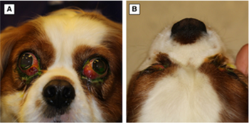 heterogeneous
contrast enhancement (18/24). In the computed tomography studies,
the zygomatic glands were all hyperattenuating and contrast
enhancing. ... Case 9: Frontal (A) and dorsoventral (B) views of a
4-year-1-month-old male neutered Cavalier King Charles
Spaniel at presentation (Case 9). Note the marked bilateral
exophthalmos, dorsolateral globe deviation, third eyelid protrusion
and conjunctival hyperaemia. There was a paraxial ventral
superficial corneal ulcer in both eyes due to the corneal exposure
(lagophthalmos), which have been stained with fluorescein dye. (C)
Post-contrast T1W and (D) T2-weighted (T2W) transverse MRI of Case
9. Note the enlarged zygomatic glands with a patchy contrast uptake
on the T1-W image and diffusely hyperintense on T2W. The left
masseter (yellow arrows) and temporalis (red arrows) muscles reveal
streaky contrast enhancement. ... Treatment included systemic
antimicrobial (18/20), anti-inflammatory (14/20) and supportive
treatment (16/20). Clinical signs improved in 16/20 dogs; however,
4/20 dogs were euthanised due to severe systemic disease. Clinical
Significance: Bilateral zygomatic sialadenitis is frequently
associated with systemic disease in dogs. Clinical signs generally
improve with systemic antimicrobial, anti-inflammatory and
supportive treatment.
heterogeneous
contrast enhancement (18/24). In the computed tomography studies,
the zygomatic glands were all hyperattenuating and contrast
enhancing. ... Case 9: Frontal (A) and dorsoventral (B) views of a
4-year-1-month-old male neutered Cavalier King Charles
Spaniel at presentation (Case 9). Note the marked bilateral
exophthalmos, dorsolateral globe deviation, third eyelid protrusion
and conjunctival hyperaemia. There was a paraxial ventral
superficial corneal ulcer in both eyes due to the corneal exposure
(lagophthalmos), which have been stained with fluorescein dye. (C)
Post-contrast T1W and (D) T2-weighted (T2W) transverse MRI of Case
9. Note the enlarged zygomatic glands with a patchy contrast uptake
on the T1-W image and diffusely hyperintense on T2W. The left
masseter (yellow arrows) and temporalis (red arrows) muscles reveal
streaky contrast enhancement. ... Treatment included systemic
antimicrobial (18/20), anti-inflammatory (14/20) and supportive
treatment (16/20). Clinical signs improved in 16/20 dogs; however,
4/20 dogs were euthanised due to severe systemic disease. Clinical
Significance: Bilateral zygomatic sialadenitis is frequently
associated with systemic disease in dogs. Clinical signs generally
improve with systemic antimicrobial, anti-inflammatory and
supportive treatment.
CONNECT WITH US