Liver / Hepatic Disorders of the
Cavalier King Charles Spaniel
-
 What
It Is
What
It Is - List of Disorders
- Symptoms
- Diagnosis
- Treatment
- What You Can Do
- Research News
- Related Links
- Veterinary Resources
The predominance of disorders of the canine liver appear to vary depending upon the breed. The most common liver disorder in dogs is chronic hepatitis. Overall, the cavalier King Charles spaniel does not stand out as having a higher incidence of any form of hepatitis, hepatic cancer, of other severe disorders involving the liver.
Below is a listing of liver disorders of which the CKCS tends to be predisposed.
RETURN TO TOP
What It Is
 The
dog's liver is a large organ and gland located in the abdomen beneath
the heart. It has several important functions, including filtering
toxins from the blood, regulating the amount of blood in the system,
making bile, which is a fluid that aids the digestion of food, and
storing vitamins and energy sources for later use. It is a gland as well
as an organ, becaue it produces bile and hormones and proteins for the
rest of the body.
The
dog's liver is a large organ and gland located in the abdomen beneath
the heart. It has several important functions, including filtering
toxins from the blood, regulating the amount of blood in the system,
making bile, which is a fluid that aids the digestion of food, and
storing vitamins and energy sources for later use. It is a gland as well
as an organ, becaue it produces bile and hormones and proteins for the
rest of the body.
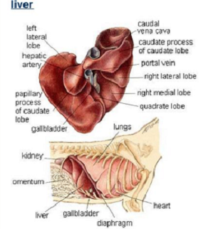
The liver consists of four lobes. Two large lobes (right lobe and left lobe) are made up of blood vessels and two small lobes (caudate lobe and quadrate lobe -- lobules) connect with bile ducts that transport bile to the small intestine. Issues and disorders related to the issue are called hepatic conditions.
Bile is a yellow fluid which includes fat and cholesterol and which is secreted through its bile duct by the liver into the small intestine to help breakdown fats in the dog's ingested food. The bile enables the vitamins and other nutrients to be absorbed into the bloodstream and also removes certain wastes.
The hepatobiliary system is a combination of the liver and bile ducts, and includes the gallbladder, a pear-shaped sac located between the two lobes of the liver, which stores bile. Read more about the cavalier's gallbladder here.
RETURN TO TOP
List of Disorders
The disorders include:
- Hepatic lesions
- Hepatic mineralization
- Hepatocutaneous syndrome
- Portal hypertension
- Portosystemic (liver) shunt
- Copper associated hepatopathy (CuAH)
- Xylitol toxicosis
Hepatic lesions
Liver lesions are groups of abnormal cells in the dog's liver. They occur as a result of any of a variety of hepatic diseases which involve inflammation of the liver. Canine hepatic diseases are divided into two main types: primary and secondary. "Primary" means that the liver is directly affected by the disorder and consists of chronic hepatitis, acute hepatitis, fibrosis, cirrhosis, and neoplasia. "Secondary" means that the disorder is ancillary to some other bodily disorder and consist of hepatic congestion, hepatic vacuolation, and reactive hepatitis.
In a January 2016 article about the prevalence of hepatic lesions in cavalier King Charles spaniels, the livers of 54 deceased CKCSs were examined. Of those, 13 were found to have no liver disorders, and the remaining 41 had hepatic lesions associated with a combined total of 7 liver disorders. Only two of the cavaliers had been diagnosed with hepatic disorders prior to their deaths. The breakdown of hepatic disorders appears on the study's Table 3 below.
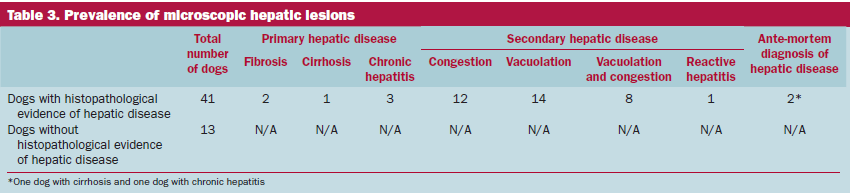
Secondary hepatic lesions were detected in 64.8% of the cavaliers in the study, while only 11.1% of them had primary hepatic lesions. The investigators stated:
"Hepatic lesions were commonly detected in this study, but the majority of cases were diagnosed as secondary to other diseases. For instance, congestion was assumed to be related to chronic heart disease. Although this supposition was not supported by the relative risk statistics, the sample size was small, and the power to detect these associations was weak. The finding of hepatocyte vacuolation is likely to be a secondary reactive change of the liver to a variety of other diseases."
In a January 2025 article, French clinicians reported the case study of a 12-year-old female cavalier found to have a very large cystic hepatic mass attached to its caudate liver lobe, one of the four lobes of the liver. The previous 15 months, the CKCS had a history of abdominal distension. Computed tomography (CT) showed the large mass. The caudate liver lobe was removed surgically, and further examination showed that the mass had necrotic tissue and signs of bleeding and remodeling. They diagnosed a cystic lesion but with pre-tumoral cells. A month later, they found no recurrence of the lesion.
RETURN TO TOP
Hepatic mineralization
Hepatic mineralization describes the appearance of mineral "opacities" in any of a variety of patterns in the liver in x-rays. In a December 2017 article, its researchers assessed the prevalence and clinical significance of linear branching mineralization of dogs' livers on x-rays from one referral veterinary hospital over a period off 30 years. They report that of 17 dogs with branching mineralization in their livers, 7 (41%) were cavalier King Charles spaniels. While they found that the prevalence of branching hepatic mineralisation in dogs appears to be very low, the CKCS was "over-represented" in the study and have an increased prevalence compared with other breeds or crossbreed dogs, since they accounted for only 5.2% of dogs seen at the hospital and only 4.1% of x-rays which included the liver.
RETURN TO TOP
Hepatocutaneous syndrome (HCS)
Hepatocutaneous syndrome (HCS) produces lesions on the surface of the
liver, giving it a "honeycomb"
appearance on ultrasound images.
It usually is accompanied by a skin disorder, superficial
necrolytic dermatitis (SND), which is discussed on our
Skin Disorder webpage. HCS
is a progressive disorder which usually is fatal. HCS is marked by
extremely low plasma amino acids (hypoaminoacidemia) and elevated
concentrations of liver enzymes, particularly serum alka ne phosphatase.
Cavaliers occassionally have been diagnosed with HCS (see this
January 2016 article and this
January 2022 article and this
January 2023 article), but overall it is not a common disorder.
Diabetes mellitus reportedly tends to develop
during the later stages of HCS.
RETURN TO TOP
Portal hypertension
The portal vein is the source of blood which flows into the liver from the gastrointestinal tract, which includes the pancreas, gallbladder, and spleen. Portal hypertension (PH) occurs if the blood pressure in the portal vein reaches a high level (e.g., 10 mm Hg) . This type of high blood pressure may be due to any of several causes, particularly increased blood flow to the liver or increased resistance to that flow (such as due to a clot or narrowing of the vessel). PH may be the result of shunts whereby the excess blood is diverted from the arteries serving the liver. Other causes include pre-existing illnesses, such as pancreatitis, cancer, other liver disorders, or complications following surgical repair of portosystemic shunts.
While this form of hypertension is not commonly reported in cavaliers, it has been found to occur in the breed occasionally. See this May 2008 article, this January 2020 article, this September 2022 article, and this February 2023 article.
RETURN TO TOP
Portosystemic (liver) shunt
In a normal dog, blood from the gastrointestinal tract enters the portal vein, which then takes blood to the liver, which then metabolizes and detoxifies this blood before sending it back into the circulatory system to the heart. A portosystemic shunt is a blood vessel present at the fetal stage, carrying toxified blood directly from the gastrointestinal tract to the heart, bypassing the liver and shunting the blood directly into the circulatory system. These shunts are classified as either intrahepatic (inside the liver) and extrahepatic (outside the liver) shunts. An intrahepatic portosystemic shunt (IHPSS) represents a normal embryologic shunt which normally closes at birth, allowing the liver to take over its filtering, storage, and production functions. However, in some cases the intrahepatic shunt does not close down properly and the liver is unable to grow or function properly. An extrahepatic portosystemic shunt (EHPSS) is an abnormal embryonic connection between to venous systems, which completely bypasses the liver. Most portosystemic shunts are congenital, but in some cases, shunts may be acquired due to another problem with the liver, such as portal hypertension.
Yorkshire terriers and Cairn terriers reportedly appear to have an inherited basis for EHPSS. Breeds which are predisposed to EHPSS are Jack Russell terriers, Dachshunds, Miniature schnauzers, and Maltese. EHPSS have been observed mainly in small breeds. Cavaliers do not appear to be predisposed to EHPSS, but some CKCSs have been diagnosed with the disorder. See this July 2012 article and this December 2015 article and this November 2017 article for additional information regarding cavaliers with this condition.
RETURN TO TOP
Copper associated hepatopathy (CuAH)
Bottom Line: Because cavaliers are predisposed to copper associated hepatopathy (CuAH), and the AAFCO refuses to recommend limits to the maximum amounts of copper added to commercial dog foods in the USA, CKCS owmers should be hesitant to feed their dogs commercial dog foods without being assured that the copper concentrations are low, such as in the range of 2.1 mg/1,000 kcal ME.
 The
metal copper (Cu) is an essential mineral in dogs' diets. It is a
component of enzymes which support respiration, for the links between
collagen and elastin, which maintains growth of bones and connective
tissues, and several other aids to cell functions.
The
metal copper (Cu) is an essential mineral in dogs' diets. It is a
component of enzymes which support respiration, for the links between
collagen and elastin, which maintains growth of bones and connective
tissues, and several other aids to cell functions.
Copper is a natural ingredient in organ meats, shellfish, sweet potatoes, beans, nuts, mushrooms, leafy greens, and dairy whey. Since 1998, the Association of American Feed Control Officials (AAFCO) has recommended that minimum copper concentrations in dry foods be 2.1 mg/1,000 kcal ME. Because most commercial dog foods do not provide enough natural ingredients containing copper, especially dry foods after processing, inorganic synthetic copper is added to those ingredients as a supplement, usually in the form of copper sulfate or copper carbonate.
Of all minerals consumed by dogs, copper is the most likely to be over-supplemented, resulting in copper associated hepatopathy (CuAH). The cavalier breed has been found to be predisposed to CuAH. CuAH describes excessive quantities of copper in the dog's liver. Copper normally is processed by the liver into bile by certain proteins. When the dog's liver is unable to convert copper, it accumulates in the liver and causes inflammation, scarring of the liver, and death of liver cells. Symptoms of CuAH rarely are evident before permanent damage to the liver's cells occur.
The primary cause of CuAH in cavaliers and other predisposed breeds is excessive quantities of copper found in many commercial dry dog foods. Since the AAFCO recommended that minimum copper concentrations in dry foods be increased to 2.1 mg/1,000 kcal ME, investigators have reported finding that the median concentration of copper in maintenance commercial dry foods was 4.4 mg/1,000 kcal ME, with a range from 2.3 to 9.0 mg/1,000 kcal ME. See this 2013 article.
In a February 2021 article, a panel of veterinary nutritionists and hepatic specialists reported observing "Over the past 15 to 20 years, we have seen what we believe to be an increased incidence of copper-associated hepatopathy in dogs." They suggested that "The onset of this increase appears to have coincided with a change in the type of copper used in premixes added to commercial dog foods." They warned that the AAFCO's "current recommendations for copper content in adult maintenance canine diets are too high and may exceed the upper tolerability limit for some dogs, resulting in hepatic disease."
In a November 2022 article, a team of Colorado State University researchers studied dogs diagnosed with "copper associated hepatopathy" (CuAH), a build-up of copper in the liver due to the liver's inability to fully process copper ingested by the dogs in their food. The researchers examined the concentrations of copper in the livers ("hepatic Cu") of 1,490 dogs in the Colorado State University Diagnostic Veterinary Laboratory database between 2010 and 2020. They report finding that the cavalier King Charles spaniel ranked sixth (after Doberman pinscher, corgi, Dalmatian, Labrador retriever, and West Highland white terrier) among all breeds with abnormally high hepatic Cu. They describe the CKCS as predisposed to CuAH. Abnormally high hepatic Cu symptoms are severe inflammation (necroinflammatory disease) of the liver, necrosis, and apoptosis (self-destruction of cells). They attribute the CuAH to excessively high quantities of copper in commercial dry dog foods. They concluded:
"Therefore, high Cu in commercial dog foods and the connection to abnormally high hepatic Cu warrants reconsideration of the current regulations for dietary Cu."
The AAFCO guidelines for minimum and maximum nutrient contents in commercial dog foods, has no limit on the maximum amount of copper that may be included in dog foods. As a result of the two veterinary studies described above, warning about abnormally high numbers of cases of hepatic Cu in cavaliers and other predisposed breeds, the AAFCO conducted a study to determine if Cu quantities in dog foods should be capped. Notwithstanding these calls for recommending to commercial dog food companies that the maximum amount of Cu in their foods be limited, in March 2023, the AAFCO announced its decision to not recommend any limit in the amount of Cu in commercial dog foods. It stated:
"Until such time as science definitively shows additional controls or restrictions are needed, AAFCO feels that recommendations for Cu concentration in foods for normal dogs are appropriately and sufficiently regulated at present." (Emphasis added.)
The term "normal dogs" is important to note, because the veterinary journal articles summarized in this section demonstrate that cavaliers are not normal dogs in terms of the processing of Cu in their diets. The high quantities of copper which the AAFCO authorizes may be tolerable for "normal" dogs, but can be highly toxic for cavaliers. Contrary to the AAFCO, the regulatory agencies in European Union (EU) countries have placed limits on the maximum amounts of copper which may be included in commercial dog foods.
In a January 2023 article, a team of Angell Animal Medical Center (Boston, Mass.) clinicians diagnosed a cavalier with superficial necrolytic dermatitis and successfully treated him with copper chelation therapy. The dog intially had high liver enzymes. Ultimately the CKCS was diagnosed with copper-associated hepatitis. A month after the diagnosis of CuAH, the dog developed hyperkeratosis (a thickening of the outer layer of the skin) on his foot pads and ulcers on his feet, legs, and rectum. Superficial necrolytic dermatitis was diagnosed at that point. They clinicians treated the dog with the copper chelation agent penicillamine designed to remove excess copper from the blood system. This drug was successful in improving the skin lesions over a period of a few months, after which the lesions returned. The dog was switched from penicillamine to stonger copper chelation agent, trientine, followed by the addition of zinc acetate, and the skin conditions improved.
In a November 2023 article, a team of Israeli researchers and Dr. Penny J. Watson reported finding copper-associated chronic hepatitis (CuCH) in 12 cavaliers. They listed a variety of common symptoms, including decreased appetite, diarrhea, vomiting, portal hypertension, ascites, increased transamination (degradation of essential amino acids), and elevated biliary enzyme concentrations. Six of the dogs (50%) had 3 or more abnormalities in measures of liver function, including hypocholesterolemia, hypoalbuminemia, hyperbilirubinemia, decreased urea concentration and hyperammonemia. All of the dogs were homozygous negative for the COMMD1 deletion mutation reported in Bedlington terriers, but 9 of them (75%) were homozygous positive or heterozygous for the ATP7B mutation reported in Labrador retrievers. The median copper score was 3/5. They found "marked architectural distortion by regenerative nodules, severe, often bridging, fibrosis, moderate inflammatory infiltrate, biliary ductular reaction and rare necrosis." Where liver fibrosis was significant, copper accumulation was most conspicuous within regenerative nodules. They treated the dogs with copper chelation, and anti-inflammatories and antioxidants, which resulted in normalization of alanine transaminase activity in 5 of 6 dogs, including resolution of ascites and liver function abnormalities in one dog. The remaining 7 dogs died despite or without chelation therapy. They concluded:
"In conclusion, we report an apparent breed-related CuCH in CKCS in which copper seemed to be a primary trigger of CH, rather than an epiphenomenon. Histochemical staining and quantitative copper measurements were significant, there was an apparent response to chelation therapy and there was a dearth of evidence to support cholestasis/inflammation-induced HCA in these dogs. An insidious onset of disease is suspected, which can be recognised by elevations in ALT activity. Notwithstanding a normal history in some dogs, or mild, nebulous clinical signs in others, significant architectural changes, fibrosis and differential copper staining within regenerative nodules were common in many cases. All affected dogs were homozygous negative for the COMMD1 and ATP7A variants but homozygous positive or heterozygous for the ATP7B variant reported in LRs, alluding to a possible genetic breed predisposition. However, the occurrence of the ATP7B variant in the control dogs demonstrates that the ATP7B variant is not singularly causative in the pathogenesis of CuCH in CKCS. Resolution of haematological and biochemical changes and clinical signs is feasible, even in the presence of previously reported negative prognostic markers. Future studies in a larger cohort of CKCS are warranted to ascertain heritability, the significance of currently found and additional genetic mutations, and the efficacy of chelation therapy and diet."
Because cavaliers are predisposed to copper associated hepatopathy (CuAH), and the AAFCO refuses to recommend limits to the maximum amounts of copper added to commercial dog foods in the USA, CKCS owmers should be hesitant to feed their dogs commercial dog foods without being assured that the copper concentrations are low, such as in the range of 2.1 mg/1,000 kcal ME.
RETURN TO TOP
Xylitol toxicosis
In an October 2009 article, a clinician reported that a cavalier which had eaten 2 or 3 pieces of chewing gum containing xylitol subsequently developed jaundice, severe lethary, anorexiz, and vomiting over a 5-day period. Laboratory testing indicated several adnormalities indicating liver damage. Helatic lymphadenopahty was detected on abdominal ultrasound. Xylitol toxicosis was presumed. LIver support therapy was started, and the dog recovered following several weeks of treatment.
RETURN TO TOP
Symptoms
The symptoms of liver disorders tend to progress from non-specific ones (increase in thirst, vomiting, diarrhea, weight loss) during the early stages to much more specific signs, such as:
• Jaundice - yellowish color of the skin, gums, and eyes
• Enlargement of the liver, resulting in noticeable swelling of the abdomen
• Blood in urine and feces
• Blindness
• Seizures
RETURN TO TOP
Diagnosis
 Blood tests, including the liver function test, are necessary to check the levels of liver
proteins, bile acids, and enzymes. Urinalysis likewise will indicate the levels.
Blood tests, including the liver function test, are necessary to check the levels of liver
proteins, bile acids, and enzymes. Urinalysis likewise will indicate the levels.
The most useful measurements are of the serum bile acids (SBA) level and albumin (Alb) level. Dropping Alb often is the first sign of liver damage. Elevated SBA likewise indicate liver dysfunction.
The major enzymes associated with hepatic injury or altered function include ALP (alkaline phosphatase), ALT (alanine transaminase) , and AST (aspartate transaminase).
Alanine aminotransaminase (ALT) , sometimes shortened to alanine transaminase, is an enzyme found in the liver that in involved in converting proteins into energy for the liver cells. When the liver is damaged, ALT is released into the bloodstream and ALT levels increase. Steroids may cause high ALT values, indicating liver damage due to the steroids. The test for ALT levels is referred to as SGPT (serum glutamic-pyruvic transaminase).
Elevated ALT does not necessarily mean liver damage. When there are no other liver-related clinical signs observed, elevated ALT may relate to a gastrointestinal issue, which could be chronic or temporaary, such as gastroenteritis.
Aspartate transaminase (AST) is an enzyme that helps the body break down amino acids. AST usually is present in the blood at low levels. An increase in AST levels may mean liver damage, liver disease, or muscle damage, including the heart, unrelated to the liver. The AST test is referred to as SGOT (glutamic-oxaloacetic transaminase).
Alkaline phosphatase (ALP or ALKP) is an enzyme that's found throughout the dog's body, but particularly in the liver, intestines, and bones. ALP blood tests measure the level of ALP in the blood that comes from the liver and bones. The range of normal levels of ALP in the blood are from 15 to 140 IU/L. High levels of ALP produced in the liver and detected in the blood may indicate liver disease or certain bone disorders. Witness Lepto is a brand name (by Zoetis) antibody test to detect IgM antibodies against Leptospra. Elevated ALP does not necessarily mean liver failure.
MicroRNAs (miRNAs), mainly miR-122, have been found to be involved in various canine hepatic diseases. They are non-coding RNAs which regulate post-transcriptional gene expression. In dogs, miR-122 has been found to have greater sensitivity than ALT in identifying hepatocellular damage. MiR-122 also aids in differentiating between hepatic parenchymal and biliary diseases. However, miR-122 upregulation also occurs in hepatocellular damage unrelated to copper overload, which makes it unsuitable as a biomarker for elevated hepatic copper. See this November 2016 article and this June 2021 article. In this March 2025 article, researchers found that cfa-miR-30b was significantly upregulated in Labrador retrievers with high hepatic copper levels. Ironically, among cavaliers, those with upregulated cfa-miR-30b showed less mitral valve disease (MVD) progression than those with lower cfa-miR-30b levels. Another study found that CKCSs in Stage B1 of MVD had elevated cfa-miR-30b-5p levels. This suggests a possible relationship between cavaliers' predisposition to both hepatic copper accumulation and MVD.
Other liver tests may include: cholesterol (Chol), total bilirubin (Tbil), and gamma-glutamyl transferase (GGT).
X-rays (radiographs) of the liver and surrounding ograns, as well as ultrasound scans are appropriate, to determine if the liver is enlarged and if cysts or tumors appear to be present.
Computed tomography angiography (CTA) is a combination of a CT scan with a contrast dye injected into a vein, to highlight blood vessels and organs, the liver in this case. CTA is used to diagnose intrahepatic portosystemic shunts (IHPSS).
Biopsies of the liver may be necessary, depending upon what is observed in the x-rays and ultrasound scans.
RETURN TO TOP
Treatment
Standard forms of treatment of liver disorders include intravenous fluid therapy to treat or avoid dehydration, and medications to control symptoms, such as vomiting and inflammation. Vitamin K (potassium) may be injected.
 The dog's glutathione, an antioxidant which is produced by the liver,
may have to be replaced or supplemented. Medications include
SAMe (S-Adenosylmethionine) and
milk thistle (silybin).
Denamarin is a combination of
SAMe and silybin. Also, ursodiol (Actigall, Urso, Ursofalk),
a choleretic bile acid, may be administered.
The dog's glutathione, an antioxidant which is produced by the liver,
may have to be replaced or supplemented. Medications include
SAMe (S-Adenosylmethionine) and
milk thistle (silybin).
Denamarin is a combination of
SAMe and silybin. Also, ursodiol (Actigall, Urso, Ursofalk),
a choleretic bile acid, may be administered.
Standard Process offers its Canine Hepatic Support as a supplement to suppor the dog's liver.
In most cases, high quality protein sources in food are recommended.
In an August 2024 article, 31 dogs diagnosed with chronic hepatobiliary disease, including 3 cavaliers, were studied to determine the efficacy of treating affected dogs with probiotics (synbiotic complex) for 4 to 6 weeks showed a significant reduction in ALT levels and clinical improvement in gastrointestinal signs. The brand of probiotic in this study was Florentero by Candioli.
Holistic treatments, such as acupuncture and Tu-Na (Chinese massage) modalities, can be helpful in treating and even healing liver disorders.
Surgery may be necessary to remove cysts or tumors.
Copper chelation agents, for removing copper and other heavy metals from the dog's blood system, include penicillamine (d-Penicillamine) and trientine. d-Penicillamine is the sulfhydryl-containing amino acid cysteine but substituted with 2 methyl groups. It reportedly significantly increases urinary excretion of copper, but with potentially serious side effects, including neurological symptoms and hypothyroidism, proteinuria, leukopenia, and thrombocytopenia. Alternative copper chelation agents include the previously mentioned trientine and zinc.
Dry dog foods with high dietary copper (Cu) content should be avoided, as should most organ meats -- especially liver -- except for beef heart. Foods high in zinc -- which binds with copper -- and eggs, dairy, seafoods, and beef heart are recommended ingredients.
RETURN TO TOP
What You Can Do
 Consider adding
milk thistle
(50 mg. per 10 lbs. body weight) as a supplement to your cavaier's daily
diet once any liver disorder is suspected. Milk thistle is a flower in the aster family. It contains an active
ingredient called silymarin, an antioxidant which supports the bodily
systems that control inflammation and stimulate new cell production.
Silymarin is a combination of three biochemicals found in milk thistle --
silychristine, silydanin, and silybin (silibinin).
Consider adding
milk thistle
(50 mg. per 10 lbs. body weight) as a supplement to your cavaier's daily
diet once any liver disorder is suspected. Milk thistle is a flower in the aster family. It contains an active
ingredient called silymarin, an antioxidant which supports the bodily
systems that control inflammation and stimulate new cell production.
Silymarin is a combination of three biochemicals found in milk thistle --
silychristine, silydanin, and silybin (silibinin).
In a June 2021 article, a team of Polish researchers studied the effect of silybin on nutrients digestibility, liver function indices, and the overall health status in 18 healthy laboratory beagles and then in 15 client-owned dogs diagnosed with idiopathic hepatic disorders. They reported finding that:
"[I]n healthy dogs, supplementation with silybin, at 12.75 mg per 10 kg BW, or with a commercial hepatoprotectant containing silybin, at the same dose, does not interfere with the nutrient's digestion, and subsequently exerts no detrimental effect on liver function indices, health, or blood parameters."
In dogs with liver disorders, they found that:
"[S]upplementation with commercial hepatoprotectant containing silybin, at a dose of 12.75 mg per 10 kg BW [body weight], decreased the activity of serum liver markers, which hence was accompanied by a decrease in the concentration of liver-specific miRNA molecules (mainly miR-122). Liver function was thus improved. Overall, silybin supplementation has no adverse impact on healthy dogs and supports liver function in dogs with hepatopathies."
MiR-122 (the blood molecule MicroRNA-122), has been found in an August 2018 study, to be a biomarker for liver injury in dogs, with very high levels of miR-122 in dogs diagnosed with liver disorders. Those researchers concluded that, "Liver disease can be sensitively and specifically diagnosed in dogs by measurement of miR-122."
RETURN TO TOP
Research News
January 2025:
Liver lobe of a cavalier is removed due to a very large hepatic
lesion.
 In
a
January 2025 article, French clinicians Theo Corbarieu (right),
Renaud Jossier, and Stephanie Claeys report the case study of a
12-year-old female cavalier King Charles spaniel found to have a very
large cystic hepatic mass attached to its caudate liver lobe, one of the
four lobes of the liver. The previous 15 months, the CKCS had a history
of abdominal distension. Computed tomography (CT) showed the large mass.
The caudate liver lobe was removed surgically, and further examination
showed that the mass had necrotic tissue and signs of bleeding and
remodeling. They diagnosed a cystic lesion but with pre-tumoral cells. A
month later, they found no recurrence of the lesion.
In
a
January 2025 article, French clinicians Theo Corbarieu (right),
Renaud Jossier, and Stephanie Claeys report the case study of a
12-year-old female cavalier King Charles spaniel found to have a very
large cystic hepatic mass attached to its caudate liver lobe, one of the
four lobes of the liver. The previous 15 months, the CKCS had a history
of abdominal distension. Computed tomography (CT) showed the large mass.
The caudate liver lobe was removed surgically, and further examination
showed that the mass had necrotic tissue and signs of bleeding and
remodeling. They diagnosed a cystic lesion but with pre-tumoral cells. A
month later, they found no recurrence of the lesion.
November 2023:
Copper-associated chronic hepatitis is found in cavalier King
Charles spaniels.
 In a
November 2023 article, a
team of Israeli researchers (Ran Nivy [right], S. Kuzi, I. Gajanayake, A.
Berkowitz) and Dr. Penny J. Watson reported finding copper-associated
chronic hepatitis (CuCH) in 12 cavalier King Charles spaniels. They listed a
variety of common symptoms, including decreased appetite, diarrhea,
vomiting, portal hypertension, ascites, increased transamination
(degradation of essential amino acids), and elevated biliary enzyme
concentrations. Six of the dogs (50%) had 3 or more abnormalities in
measures of liver function, including hypocholesterolemia,
hypoalbuminemia, hyperbilirubinemia, decreased urea concentration and
hyperammonemia. All of the dogs were homozygous negative for the COMMD1
deletion mutation reported in Bedlington terriers, but 9 of them (75%)
were homozygous positive or heterozygous for the ATP7B mutation reported
in Labrador retrievers. The median copper score was 3/5. They found
"marked architectural distortion by regenerative nodules, severe, often
bridging, fibrosis, moderate inflammatory infiltrate, biliary ductular
reaction and rare necrosis." Where liver fibrosis was significant,
copper accumulation was most conspicuous within regenerative nodules.
They treated the dogs with copper chelation, and anti-inflammatories and
antioxidants, which resulted in normalization of alanine transaminase
activity in 5 of 6 dogs, including resolution of ascites and liver
function abnormalities in one dog. The remaining 7 dogs died despite or
without chelation therapy. They concluded:
In a
November 2023 article, a
team of Israeli researchers (Ran Nivy [right], S. Kuzi, I. Gajanayake, A.
Berkowitz) and Dr. Penny J. Watson reported finding copper-associated
chronic hepatitis (CuCH) in 12 cavalier King Charles spaniels. They listed a
variety of common symptoms, including decreased appetite, diarrhea,
vomiting, portal hypertension, ascites, increased transamination
(degradation of essential amino acids), and elevated biliary enzyme
concentrations. Six of the dogs (50%) had 3 or more abnormalities in
measures of liver function, including hypocholesterolemia,
hypoalbuminemia, hyperbilirubinemia, decreased urea concentration and
hyperammonemia. All of the dogs were homozygous negative for the COMMD1
deletion mutation reported in Bedlington terriers, but 9 of them (75%)
were homozygous positive or heterozygous for the ATP7B mutation reported
in Labrador retrievers. The median copper score was 3/5. They found
"marked architectural distortion by regenerative nodules, severe, often
bridging, fibrosis, moderate inflammatory infiltrate, biliary ductular
reaction and rare necrosis." Where liver fibrosis was significant,
copper accumulation was most conspicuous within regenerative nodules.
They treated the dogs with copper chelation, and anti-inflammatories and
antioxidants, which resulted in normalization of alanine transaminase
activity in 5 of 6 dogs, including resolution of ascites and liver
function abnormalities in one dog. The remaining 7 dogs died despite or
without chelation therapy. They concluded:
"In conclusion, we report an apparent breed-related CuCH in CKCS in which copper seemed to be a primary trigger of CH, rather than an epiphenomenon. Histochemical staining and quantitative copper measurements were significant, there was an apparent response to chelation therapy and there was a dearth of evidence to support cholestasis/inflammation-induced HCA in these dogs. An insidious onset of disease is suspected, which can be recognised by elevations in ALT activity. Notwithstanding a normal history in some dogs, or mild, nebulous clinical signs in others, significant architectural changes, fibrosis and differential copper staining within regenerative nodules were common in many cases. All affected dogs were homozygous negative for the COMMD1 and ATP7A variants but homozygous positive or heterozygous for the ATP7B variant reported in LRs, alluding to a possible genetic breed predisposition. However, the occurrence of the ATP7B variant in the control dogs demonstrates that the ATP7B variant is not singularly causative in the pathogenesis of CuCH in CKCS. Resolution of haematological and biochemical changes and clinical signs is feasible, even in the presence of previously reported negative prognostic markers. Future studies in a larger cohort of CKCS are warranted to ascertain heritability, the significance of currently found and additional genetic mutations, and the efficacy of chelation therapy and diet."
January 2023:
Cavalier with copper-associated hepatitis is successfully treated
with copper chelation for necrolytic dermatitis.  In
a
January 2023 article, a team of Angell Animal Medical Center (Boston,
Mass.) clinicians (Cynthia Talbot, Shawn Kearns [right], Pamela J.
Mouser) diagnosed a cavalier King Charles spaniel with superficial
necrolytic dermatitis and successfully treated him with copper chelation
therapy. The dog intially had high liver enzymes. Ultimately the CKCS was
diagnosed with copper-associated hepatitis. Cavaliers as a breed have been
found to be predisposed to copper associated hepatopahty (CAH). CAH
describes excessive quantities of the metal copper (Cu) in the dog's liver.
Cavaliers appear to be less capable of processing copper by the liver into
bile. When the dog's liver is unable to convert copper, it accumulates in
the liver and causes inflammation, scarring of the liver, and death of liver
cells. A month after the diagnosis of CAH, the dog developed hyperkeratosis
(a thickening of the outer layer of the skin) on his foot pads and ulcers on
his feet, legs, and rectum. Superficial necrolytic dermatitis was diagnosed
at that point. They clinicians treated the dog with the copper chelation
agent penicillamine designed to remove excess copper from the blood system.
This drug was successful in improving the skin lesions over a period of a
few months, after which the lesions returned. The dog was switched from
penicillamine to stonger copper chelation agent, trientine, followed by the
addition of zinc acetate, and the skin conditions improved.
In
a
January 2023 article, a team of Angell Animal Medical Center (Boston,
Mass.) clinicians (Cynthia Talbot, Shawn Kearns [right], Pamela J.
Mouser) diagnosed a cavalier King Charles spaniel with superficial
necrolytic dermatitis and successfully treated him with copper chelation
therapy. The dog intially had high liver enzymes. Ultimately the CKCS was
diagnosed with copper-associated hepatitis. Cavaliers as a breed have been
found to be predisposed to copper associated hepatopahty (CAH). CAH
describes excessive quantities of the metal copper (Cu) in the dog's liver.
Cavaliers appear to be less capable of processing copper by the liver into
bile. When the dog's liver is unable to convert copper, it accumulates in
the liver and causes inflammation, scarring of the liver, and death of liver
cells. A month after the diagnosis of CAH, the dog developed hyperkeratosis
(a thickening of the outer layer of the skin) on his foot pads and ulcers on
his feet, legs, and rectum. Superficial necrolytic dermatitis was diagnosed
at that point. They clinicians treated the dog with the copper chelation
agent penicillamine designed to remove excess copper from the blood system.
This drug was successful in improving the skin lesions over a period of a
few months, after which the lesions returned. The dog was switched from
penicillamine to stonger copper chelation agent, trientine, followed by the
addition of zinc acetate, and the skin conditions improved.
November 2022:
Copper-associated chronic hepatitis is found in cavalier King
Charles spaniels.
In a
September 2022 abstract presented at the 32d European College of
Veterinary Internal Medicine - Companion Animals (ECVIM-CA) Congress, a
team of Israeli researchers (Ran Nivy [right], S. Kuzi, I. Gajanayake, A.
Berkowitz) and Dr. Penny J. Watson reported finding copper-associated
chronic hepatitis in 12 cavalier King Charles spaniels. They listed a
variety of common symptoms, including decreased appetite, diarrhea,
vomiting, portal hypertension, ascites, increased transamination
(degradation of essential amino acids), and elevated biliary enzyme
concentrations. Six of the dogs (50%) had 3 or more abnormalities in
measures of liver function, including hypocholesterolemia,
hypoalbuminemia, hyperbilirubinemia, decreased urea concentration and
hyperammonemia. All of the dogs were homozygous negative for the COMMD1
deletion mutation reported in Bedlington terriers, but 9 of them (75%)
were homozygous positive or heterozygous for the ATP7B mutation reported
in Labrador retrievers. The median copper score was 3/5. They found
"marked architectural distortion by regenerative nodules, severe, often
bridging, fibrosis, moderate inflammatory infiltrate, biliary ductular
reaction and rare necrosis." Where liver fibrosis was significant,
copper accumulation was most conspicuous within regenerative nodules.
They treated the dogs with copper chelation, and anti-inflammatories and
antioxidants, which resulted in normalization of alanine transaminase
activity in 5 of 6 dogs, including resolution of ascites and liver
function abnormalities in one dog. The remaining 7 dogs died despite or
without chelation therapy. They concluded:
"Considering the findings herein, CuCH should be considered in CKCS with suspected liver disease. Long-term prognosis, albeit based on a small number of dogs, seems favorable in dogs receiving chelation therapy, notwithstanding the presence of previously reported negative prognostic markers."
November 2022:
Cavaliers are found predisposed to copper associated
hepatopathy, due to increases in copper content in commercial dry dog
foods.
 In
a
November 2022 article, a team of Colorado State University
researchers (Tarini Vedantham Ullal, Steven Lakin, Brooke Gallagher,
Nick Sbardellati, Zaid Abdo, David C. Twedt [right]) studied
dogs diagnosed with "copper associated hepatopathy" (CAH), a build-up of
copper in the liver due to the liver's inability to fully process copper
ingested by the dogs in their food. The researchers examined the
concentrations of copper in the livers ("hepatic Cu") of 1,490 dogs in
the Colorado State University Diagnostic Veterinary Laboratory database
between 2010 and 2020. They report finding that the cavalier King
Charles spaniel ranked sixth (after Doberman pinscher, corgi, Dalmatian,
Labrador retriever, and West Highland white terrier) among all breeds
with abnormally high hepatic Cu. They describe the CKCS as predisposed
to CAH. Abnormally high hepatic Cu symptoms are severe inflammation
(necroinflammatory disease) of the liver, necrosis, and apoptosis
(self-destruction of cells). They attribute the CAH to excessively high
quantities of copper in commercial dry dog foods. They observe that,
beginning in 1998, the Association of American Feed Control Officials
(AAFCO) recommended that minimum copper concentrations in dry foods be
2.1 mg/1,000 kcal ME. A
prior study reported finding that the median
concentration of copper in maintenance commercial dry foods was 4.4
mg/1,000 kcal ME, with a range from 2.3 to 9.0 mg/1,000 kcal ME. The
investigators in the present study conclude:
In
a
November 2022 article, a team of Colorado State University
researchers (Tarini Vedantham Ullal, Steven Lakin, Brooke Gallagher,
Nick Sbardellati, Zaid Abdo, David C. Twedt [right]) studied
dogs diagnosed with "copper associated hepatopathy" (CAH), a build-up of
copper in the liver due to the liver's inability to fully process copper
ingested by the dogs in their food. The researchers examined the
concentrations of copper in the livers ("hepatic Cu") of 1,490 dogs in
the Colorado State University Diagnostic Veterinary Laboratory database
between 2010 and 2020. They report finding that the cavalier King
Charles spaniel ranked sixth (after Doberman pinscher, corgi, Dalmatian,
Labrador retriever, and West Highland white terrier) among all breeds
with abnormally high hepatic Cu. They describe the CKCS as predisposed
to CAH. Abnormally high hepatic Cu symptoms are severe inflammation
(necroinflammatory disease) of the liver, necrosis, and apoptosis
(self-destruction of cells). They attribute the CAH to excessively high
quantities of copper in commercial dry dog foods. They observe that,
beginning in 1998, the Association of American Feed Control Officials
(AAFCO) recommended that minimum copper concentrations in dry foods be
2.1 mg/1,000 kcal ME. A
prior study reported finding that the median
concentration of copper in maintenance commercial dry foods was 4.4
mg/1,000 kcal ME, with a range from 2.3 to 9.0 mg/1,000 kcal ME. The
investigators in the present study conclude:
"Therefore, high Cu in commercial dog foods and the connection to abnormally high hepatic Cu warrants reconsideration of the current regulations for dietary Cu."
June 2021:
Silybin in milk thistle improved liver function in dogs with
hepatopathies, in Polish study.
 In a
June 2021 article, a team of Polish researchers (Maciej Gogulski, Adam
Cieślak, Julia Grabska, Marie Ardois, Małgorzata Pomorska-Mól, Paweł A.
Kołodziejski, Kacper Libera, Viola Strompfová, Małgorzata Szumacher-Strabel
[right]) studied the effect of
silybin on nutrients digestibility, liver function indices, and the overall
health status in 18 healthy laboratory beagles and then in 15 client-owned
dogs diagnosed with idiopathic hepatic disorders. They reported finding
that:
In a
June 2021 article, a team of Polish researchers (Maciej Gogulski, Adam
Cieślak, Julia Grabska, Marie Ardois, Małgorzata Pomorska-Mól, Paweł A.
Kołodziejski, Kacper Libera, Viola Strompfová, Małgorzata Szumacher-Strabel
[right]) studied the effect of
silybin on nutrients digestibility, liver function indices, and the overall
health status in 18 healthy laboratory beagles and then in 15 client-owned
dogs diagnosed with idiopathic hepatic disorders. They reported finding
that:
"[I]n healthy dogs, supplementation with silybin, at 12.75 mg per 10 kg BW, or with a commercial hepatoprotectant containing silybin, at the same dose, does not interfere with the nutrient's digestion, and subsequently exerts no detrimental effect on liver function indices, health, or blood parameters."
In dogs with liver disorders, they found that:
"[S]upplementation with commercial hepatoprotectant containing silybin, at a dose of 12.75 mg per 10 kg BW [body weight], decreased the activity of serum liver markers, which hence was accompanied by a decrease in the concentration of liver-specific miRNA molecules (mainly miR-122). Liver function was thus improved. Overall, silybin supplementation has no adverse impact on healthy dogs and supports liver function in dogs with hepatopathies."
August 2018:
UK Researchers find blood molecule microRNA-122 is marker for
liver disease in dogs.
 In
an
August 2018 article, a team of researchers (W. Oosthuyzen, P.W.L.
Ten Berg, B. Francis, S. Campbell, V. Macklin, E. Milne, A. G. Gow, C.
Fisher, R.J. Mellanby, J.W. Dear [right]) at the University of
Edinburgh's Royal (Dick) School of Veterinary Studies examined the blood
of 250 dogs (120 healthy dogs, 100 dogs with non-liver diseases, and 30
dogs with confirmed liver disease) to determine if the blood molecule
MicroRNA-122 (miR-122) can be a biomarker for liver injury in dogs, as
it already has shown to be in humans and rodents. They found no
difference between healthy dogs and those with non-liver disease.
However, they found very high levels of miR-122 in dogs diagnosed with
liver disorders. They concluded that, "Liver disease can be sensitively
and specifically diagnosed in dogs by measurement of miR-122."
In
an
August 2018 article, a team of researchers (W. Oosthuyzen, P.W.L.
Ten Berg, B. Francis, S. Campbell, V. Macklin, E. Milne, A. G. Gow, C.
Fisher, R.J. Mellanby, J.W. Dear [right]) at the University of
Edinburgh's Royal (Dick) School of Veterinary Studies examined the blood
of 250 dogs (120 healthy dogs, 100 dogs with non-liver diseases, and 30
dogs with confirmed liver disease) to determine if the blood molecule
MicroRNA-122 (miR-122) can be a biomarker for liver injury in dogs, as
it already has shown to be in humans and rodents. They found no
difference between healthy dogs and those with non-liver disease.
However, they found very high levels of miR-122 in dogs diagnosed with
liver disorders. They concluded that, "Liver disease can be sensitively
and specifically diagnosed in dogs by measurement of miR-122."
December 2017:
Cavaliers are "over-represented" in a study of hepatic
mineralization.
 Hepatic mineralization describes the appearance of mineral
"opacities" in any of a variety of patterns in the liver in x-rays. In a
December 2017 article, its researchers (M.-A. Genain, A. Barbosa, M.
Herrtage [right], P. Watson) assessed the prevalence and
clinical significance of linear branching mineralization of dogs' livers
on x-rays from one referral veterinary hospital over a period off 30
years. They report that of 17 dogs with branching mineralization in
their livers, 7 (41%) were cavalier King Charles spaniels. While they
found that the prevalence of branching hepatic mineralisation in dogs
appears to be very low, the CKCS was "over-represented" in the study and
have an increased prevalence compared with other breeds or crossbreed
dogs, since they accounted for only 5.2% of dogs seen at the hospital
and only 4.1% of x-rays which included the liver.
Hepatic mineralization describes the appearance of mineral
"opacities" in any of a variety of patterns in the liver in x-rays. In a
December 2017 article, its researchers (M.-A. Genain, A. Barbosa, M.
Herrtage [right], P. Watson) assessed the prevalence and
clinical significance of linear branching mineralization of dogs' livers
on x-rays from one referral veterinary hospital over a period off 30
years. They report that of 17 dogs with branching mineralization in
their livers, 7 (41%) were cavalier King Charles spaniels. While they
found that the prevalence of branching hepatic mineralisation in dogs
appears to be very low, the CKCS was "over-represented" in the study and
have an increased prevalence compared with other breeds or crossbreed
dogs, since they accounted for only 5.2% of dogs seen at the hospital
and only 4.1% of x-rays which included the liver.
January 2016:
Secondary hepatic lesions were found in 64.8% of post-mortem
samples of cavaliers in UK study.
 In a
January 2016 article, UK researchers (Kent, Andrew C. C.;
Constantino-Casas, Fernando; Rusbridge, Clare; Corcoran, Brendan;
Carter, Margaret; Ledger, Tania; Watson, Penny J. [right]) searched for
pancreatic, hepatic (liver) and renal (kidney) lesions in post-mortem
samples from Cavalier King Charles Spaniels (CKCSs). Primary hepatic
lesions were present in only 11.1% of cases, but secondary hepatic
lesions were more common and were present in 64.8%. The researchers
report that cavaliers have similar rates of hepatic disease as the
general population.
In a
January 2016 article, UK researchers (Kent, Andrew C. C.;
Constantino-Casas, Fernando; Rusbridge, Clare; Corcoran, Brendan;
Carter, Margaret; Ledger, Tania; Watson, Penny J. [right]) searched for
pancreatic, hepatic (liver) and renal (kidney) lesions in post-mortem
samples from Cavalier King Charles Spaniels (CKCSs). Primary hepatic
lesions were present in only 11.1% of cases, but secondary hepatic
lesions were more common and were present in 64.8%. The researchers
report that cavaliers have similar rates of hepatic disease as the
general population.
RETURN TO TOP
Related Links
RETURN TO TOP
Veterinary Resources
Control of Canine Genetic Diseases. Padgett. G.A., Howell Book House 1998, pp. 198-199, 222.
Use of transcolonic portal scintigraphy to evaluate efficacy of cellophane banding of congenital extrahepatic portosystemic shunts in 16 dogs. B.P. Landon, L.A. Abraham, J.A. Charles. Australian Vet. J. May 2008; doi: 10.1111/j.1751-0813.2008.00278.x. Quote: Objective: To evaluate the efficacy of cellophane banding of single congenital extrahepatic portosystemic shunts in dogs using transcolonic portal scintigraphy. To investigate the portal circulation of those dogs with elevated postoperative shunt fractions to determine the cause of the persistent shunting. Further, to evaluate whether presenting signs, clinical pathology findings and liver histopathology are predictive of outcome. Design: Prospective study of 16 dogs presenting with single congenital extrahepatic portosystemic shunts [including one cavalier King Charles spaniel]. Procedure: Dogs with single extrahepatic portosystemic shunts attenuated by cellophane banding underwent portal scintigraphy and bile acids tolerance testing pre- and post-operatively. Dogs identified with elevated shunt fractions at 10 weeks post-operatively underwent mesenteric portovenography. Qualitative hepatic histopathology from all dogs was reviewed by a veterinary pathologist and assigned a semi-quantitative score to identify any abnormalities that may predict surgical outcome. Results: At 10 weeks post cellophane banding, 10 of 16 cases (63%) had normal shunt fractions, whilst six dogs (37%) had increased shunt fractions and seven dogs (44%) had increased serum bile acids. Of these dogs, mesenteric portovenography revealed incomplete closure of the shunt in three dogs (18.6%) and multiple acquired shunts in three dogs (18.6%). Liver histopathology findings were similar for all dogs, regardless of outcome. Conclusions: Cellophane banding is an efficacious method for complete gradual occlusion of single extrahepatic shunts when the shunt vessel is attenuated to ≤ 3 mm. Transcolonic portal scintigraphy is a reliable method for assessment of shunt attenuation and, unlike serum bile acids, is not influenced by other causes of liver dysfunction.
Acute hepatic necrosis following xylitol ingestion in a 2-year-old dog. Hillary Wentworth. Cornell Univ. eCommons. October 2009. Quote: A 2-year-old intact female Cavalier King Charles Spaniel presented to the triage service at the Cornell University Hospital for Animals (CUHA) with a 5-day history of icterus, severe lethargy, anorexia, and vomiting. The patient had a history of eating 2-3 pieces of xylitol-containing chewing gum 8 days before presentation to her referring veterinarian (48 hours before the onset of her clinical signs). Multiple laboratory abnormalities referable to liver damage were observed. Hepatic lymphadenopathy was detected on abdominal ultrasound. A presumptive diagnosis of xylitol toxicosis was made based on the patient's history and laboratory findings. Supportive and hepatoprotective therapy was instituted and the patient recovered after a treatment course of several weeks. Principals of the pathogenesis, diagnosis, treatment, and prognosis of canine xylitol toxicosis are discussed.
Distribution of extrahepatic congenital portosystemic shunt morphology in predisposed dog breeds. Lindsay Van den Bossche, Frank G van Steenbeek, Robert P Favier, Anne Kummeling, Peter AJ Leegwater, Jan Rothuizen. BMC Vet. Research. July 2012;8:112. Quote: "Background: An inherited basis for congenital extrahepatic portosystemic shunts (EHPSS) has been demonstrated in several small dog breeds. If in general both portocaval and porto-azygous shunts occur in breeds predisposed to portosystemic shunts then this could indicate a common genetic background. This study was performed to determine the distribution of extrahepatic portocaval and porto-azygous shunts in purebred dog populations. Results: Data of 135 client owned dogs diagnosed with EHPSS at the Faculty of Veterinary Medicine of Utrecht University from 2001 - 2010 were retrospectively analyzed. The correlation between shunt localization, sex, age, dog size and breed were studied. The study group consisted of 54 males and 81 females from 24 breeds. ... Additional breeds diagnosed with EHPSS were the Lhasa Apso, Miniature Poodle, Norfolk terrier with two cases, and single cases of a Basset Hound, Bolognese, Cavalier King Charles Spaniel, Epagneul Nain Papillon, Flat Coated Retriever, Fox terrier, Giant Spitz, Great Dane, Miniature Pinscher, Norwich terrier and Welsh terrier. ... Twenty-five percent of dogs had porto-azygous shunts and 75% had portocaval shunts. Of the dogs with porto-azygous shunts only 27% was male (P = 0.006). No significant sex difference was detected in dogs with a portocaval shunt. Both phenotypes were present in almost all breeds represented with more than six cases. Small dogs are mostly diagnosed with portocaval shunts (79%) whereas both types are detected. The age at diagnosis in dogs with porto-azygous shunts was significantly higher than that of dogs with portocaval shunts (P < 0.001). Conclusion: The remarkable similarity of phenotypic variation in many dog breeds may indicate common underlying genes responsible for EHPSS across breeds. The subtype of EHPSS could be determined by a minor genetic component or modulating factors during embryonic development."
Evaluation of calcium, phosphorus, and selected trace mineral status in commercially available dry foods formulated for dogs. Jason W. Gagne, Joseph J. Wakshlag, Sharon A. Center, Michael A. Rutzke, Raymond P. Glahn. J.A.V.M.A. September 2013; doi: 10.2460/javma.243.5.658. Quote: Objective: To evaluate concentrations of calcium, phosphorus, zinc, iron, copper, manganese, and selenium in several commercially available dry dog foods and compare these with current Association of American Feed Control Officials (AAFCO) recommendations for maintenance of healthy dogs. Design: Descriptive study. Sample--45 over-the-counter dry foods formulated for maintenance of healthy dogs (ie, maintenance foods) and 5 therapeutic dry foods formulated for dogs with hepatic or renal disease. Procedures: Mineral concentrations were measured via inductively coupled plasma mass spectrometry or inductively coupled plasma atomic emission spectroscopy and compared with AAFCO-recommended minimum and maximum values. ... With the exception of therapeutic foods, no products had copper concentrations below the AAFCO-recommended minimum of 2.1 mg/1,000 kcal ME; none exceeded the recommended maximum value. The median concentration of copper in maintenance foods was 4.4 mg/1,000 kcal ME (range, 2.3 to 9.0 mg/1,000 kcal ME). Copper concentrations in 2 therapeutic foods for dogs with hepatic disease were 1.3 and 1.7 mg/1,000 kcal ME. Considering that these diets were formulated to maintain neutral copper balance in Bedlington Terriers with copper-storage hepatopathy, maintenance food copper concentration was also compared with that standard. Copper concentrations in 29 of 45 (64%) maintenance foods were approximately 2 times the median value for these 2 therapeutic foods (1.5 mg/1,000 kcal ME), and copper concentrations in 4 maintenance foods were 3 times this value.
A Retrospective Histopathological Survey on Canine and Feline Liver Diseases at the University of Tokyo between 2006 and 2012. Naoki Hirose, Kazuyuki Uchida, Hideyuki Kanemoto, Koichi Ohno, James K. Chambers, Hiroyuki Nakayama. J. Vet. Med. Sci. July 2014;76(7): 1015-1020. Quote: "To determine the incidence of hepatic diseases in dogs and cats in Japan, a retrospective study was performed using data of 463 canine and 71 feline liver biopsies at the Veterinary Medical Center of the University of Tokyo. The most common canine hepatic disease was microvascular dysplasia (MVD) and occupied 29.4% of all diagnoses. This terminology might contain "real" MVD and primary portal vein hypoplasia, because these two conditions were difficult to be clearly distinguished histopathologically. Parenchymal and interstitial hepatitis and primary hepatic tumors accounted for 23.5% and 21.0% of the diagnoses, respectively. Parenchymal and interstitial hepatitis occupied 34.1% of non-proliferative canine hepatic diseases, while hepatocellular adenoma and carcinoma were 26.6% and 24.5% of roliferative hepatic diseases, respectively. Breed-specificity was seen in MVD for Yorkshire terrier, Papillon and Toy poodle, in hepatitis for Doberman pinscher and Labrador retriever, in cholangiohepatitis for American cocker spaniel, Miniature schnauzer and Pomeranian, in hepatocellular adenoma for Golden retriever and Shiba and in hepatocellular carcinoma for Shih Tzu. Among 25 cases of canine chronic hepatitis, Labrador retrievers and Doberman Pinschers ranked the first (8 cases, 32.0%) and the second (3 cases, 12.0%), respectively. Females were more susceptible than males in both breeds [Labrador retrievers (male/female=1:7) and Doberman pinschers (male/female=1:2)]. The median age of the hepatitis cases was 8 years and 7 months old. Eight of the 25 canine chronic hepatitis cases had copper deposition. Two of the 8 cases were Doberman pinschers, and 1 was Labrador retriever, Bedlington terrier, Welsh corgi, Cavalier King Charles spaniel or a mixed breed."
Lipopolysaccharide and toll-like receptor 4 in dogs with congenital portosystemic shunts. M.S. Tivers, V.J. Lipscomb, K.C. Smith, C.P.D. Wheeler-Jones, A.K. House. Vet. J. December 2015;404-2015;206(3):303-413. Quote: "Surgical attenuation of a congenital portosystemic shunt (CPSS) results in increased portal vein perfusion, liver growth and clinical improvement. Portal lipopolysaccharide (LPS) is implicated in liver regeneration via toll-like receptor (TLR) 4 mediated cytokine activation. The aim of this study was to investigate factors associated with LPS in dogs with CPSS. Plasma LPS concentrations were measured in the peripheral and portal blood using a limulus amoebocyte lysate (LAL) assay. ... We included paired peripheral and portal plasma samples from 13 CPSS dogs of the following breeds: Bichon Frise (n = 2), Labrador (n = 2), Border terrier (n = 1), Cavalier King Charles spaniel (n = 1), crossbreed (n = 1), Dogue de Bordeaux (n = 1), German shepherd dog (n = 1), Miniature Schnauzer (n = 1), Springer spaniel (n = 1), West Highland White terrier (n = 1), Yorkshire terrier (n = 1). The median age was 295 days (range, 125-1835 days). Nine dogs (69.2%) had an extrahepatic CPSS and four (30.8%) had an intrahepatic CPSS. ... LPS concentration was significantly greater in the portal blood compared to peripheral blood in dogs with CPSS (P = 0.046) and control dogs (P = 0.002). LPS concentrations in the peripheral (P = 0.012) and portal (P = 0.005) blood of dogs with CPSS were significantly greater than those of control dogs. The relative mRNA expression of cytokines and TLRs was measured in liver biopsies from dogs with CPSS using quantitative PCR. TLR4 expression significantly increased following partial CPSS attenuation (P = 0.020). TLR4 expression was significantly greater in dogs that tolerated complete CPSS attenuation (P = 0.011) and those with good portal blood flow on pre-attenuation (P = 0.004) and post-attenuation (P = 0.015) portovenography. Serum interleukin (IL)-6 concentration was measured using a canine specific ELISA and significantly increased 24 h following CPSS attenuation (P < 0.001). Portal LPS was increased in dogs with CPSS, consistent with decreased hepatic clearance. TLR4 mRNA expression was significantly associated with portal blood flow and increased following surgery. These findings support the concept that portal LPS delivery is important in the hepatic response to surgical attenuation. Serum IL-6 significantly increased following surgery, consistent with LPS stimulation via TLR4, although this increase might be non-specific."
Prevalence of pancreatic, hepatic and renal microscopic lesions in post-mortem samples from Cavalier King Charles Spaniels. Kent, Andrew C. C.; Constantino-Casas, Fernando; Rusbridge, Clare; Corcoran, Brendan; Carter, Margaret; Ledger, Tania; Watson, Penny J. J. Small Animal Practice, January 2016. Quote: "Objectives: To describe the prevalence of pancreatic, hepatic and renal microscopic lesions in post-mortem samples from Cavalier King Charles Spaniels (CKCS) presented to a UK post-mortem collection scheme. Methods: Histopathology was performed on the organs of interest and the prevalence of microscopic lesions described, this was related back to the clinical signs shown ante-mortem. Results: Evidence of chronic pancreatitis was seen in 51.9% of the cases, and age correlated with severity of disease, suggesting that chronic pancreatitis is a progressive condition. Evidence of renal lesions was present in 52.2% of cases, most commonly inflammatory disease. The rate of ante-mortem diagnosis was low for both pancreatic and renal disease, at 25% and 16.7% respectively. Primary hepatic lesions were present in only 11.1% of cases, but secondary hepatic lesions were more common and were present in 64.8%. Clinical Significance: Pancreatic and renal lesions are common in Cavalier King Charles Spaniels and clinicians should be aware of this when presented with clinical cases, they have similar rates of hepatic disease as the general population."
Morphology of splenocaval congenital portosystemic shunts in dogs and cats. R. N. White, A. T. Parry. J.S.A.P. January 2016;57(1):28-32. Quote: "Objective: To describe the anatomy of congenital portosystemic shunts involving the splenic vein communicating with the caudal vena cava at the level of the epiploic foramen. Materials and Methods: A retrospective review of a consecutive series of dogs and cats managed for congenital portosystemic shunts. Results: Ninety-eight dogs and eight cats met the inclusion criteria of a congenital portosystemic shunt involving the splenic vein communicating with the prehepatic caudal vena cava plus recorded intra-operative mesenteric portovenography or computed tomography angiography and gross observations at surgery. All cases (both dogs and cats) had a highly consistent shunt that involved a distended gastrosplenic vein that communicated with the caudal vena cava at the level of the epiploic foramen via an anomalous left gastric vein. Clinical Significance: The morphology of the shunt type described appeared to be a result of an abnormal communication between the left gastric vein and the caudal vena cava and the subsequent development of preferential blood flow through an essentially normal portal venous system. The abnormal communication (shunt) was through the left gastric vein and not the splenic vein, as might have been expected. This information may help with surgical planning in cases undergoing shunt closure surgery."
Use of Serum MicroRNAs as Biomarker for Hepatobiliary Diseases in Dogs. K. Dirksen, T. Verzijl, G.C. Grinwis, R.P. Favier, L.C. Penning, I.A. Burgener, L.J. van der Laan, H. Fieten, B. Spee. J. Vet. Intern. Med. November 2016; doi: 10.1111/jvim.14602. Quote: Background: Current biochemical indicators cannot discriminate between parenchymal, biliary, vascular, and neoplastic hepatobiliary diseases. MicroRNAs are promising new biomarkers for hepatobiliary disease in humans and dogs. Objective: To measure serum concentrations of an established group of microRNAs in dogs and to investigate their concentrations in various types of hepatobiliary diseases. Animals: Forty-six client-owned dogs with an established diagnosis of hepatobiliary disease and stored serum samples [incluidng 1 cavalier King Charles spaniel] and eleven client-owned healthy control Labrador Retrievers. Methods: Retrospective study. Medical records of dogs with parenchymal, biliary, vascular, or neoplastic hepatobiliary diseases and control dogs were reviewed. Concentrations of miR-21, miR-122, miR-126, miR-148a, miR-200c, and miR-222 were quantified in serum by real-time polymerase chain reaction. Results: No different microRNA concentrations were found in the adenoma and congenital portosystemic shunt groups. In all other diseases, miR-122 concentrations were elevated with the highest concentration in the mucocele group (267-fold, CI: 40-1,768, P < .001). In dogs with biliary diseases, miR-21 and miR-222 were only increased in dogs with mucoceles (26 fold, CI: 5-141, P = .005 and 13-fold, CI: 2-70, P = .025, respectively). Uniquely increased microRNAs were found in the hepatocellular carcinoma group (miR-200c, 35-fold increase, CI: 3-382, P = .035) and the chronic hepatitis group (miR-126, 22-fold increase, CI: 5-91, P = .002). Conclusions and Clinical Importance: A microRNA panel consisting of miR-21, miR-122, miR-126, miR-200c, and miR-222 can distinguish between parenchymal, biliary, and neoplastic hepatobiliary diseases. Serum microRNA profiling is a promising new tool that might be a valuable addition to conventional diagnostics to help diagnose various hepatobiliary diseases in dogs.
Clinical relevance of radiographic linear branching mineral opacities in the canine liver. M.-A. Genain, A. Barbosa, M. Herrtage, P. Watson. J. Sm. Anim. Pract. December 2017. DOI: 10.1111/jsap.12797. Quote: Various conditions have been reported to cause mineral opacities within the liver or biliary system in dogs, including parenchymal mineralisation or linear branching mineralisation. Hepatic parenchymal mineral opacification has been described in different patterns: a densely mineralised lesion, a curvilinear ring of calcification or large centrally located coarse mineralisation pattern or multiple or solitary foci of mineralisation located eccentrically within a complex heterogeneous mass. ... Branching mineralisation within the hepatic parenchyma of the dog has been reported occasionally in older small breed dogs and is the focus of this study. ... Objectives: To assess the prevalence, clinical significance and breed distribution of linear branching mineralisation superimposed on the hepatic radiographic silhouette in dogs. Materials and Methods: Retrospective review of radiographs or ultrasound images of dogs showing branching mineralisation in the liver. Results: Over the 30-year review period, 17 cases were identified and the mineralisation had a predominantly ventral distribution. Seven of the 17 were cavalier King Charles spaniels, and four of the total 17 dogs were diagnosed with hepatobiliary system disease. Five dogs had repeat radiographs, of which four showed no change in the pattern and one developed the pattern 6 years after being diagnosed with cholangiohepatitis. Serum calcium concentrations were normal in all patients. Liver enzymes were markedly elevated only in the dog diagnosed with cholangiohepatitis. Histology performed on three patients showed no convincing evidence of primary liver disease. Clinical Significance: ... Hepatic linear branching mineralisation is uncommon in dogs. CKCS appear to have an increased prevalence compared with other breeds or crossbreed dogs. Biopsy of the liver of affected dogs with no clinical or clinicopathological evidence of liver disease is unlikely to be helpful in these cases.
Intrahepatic congenital portosystemic shunts in dogs: short- and long-term outcome of suture attenuation. M. S. Tivers, V. J. Lipscomb, P. Bristow, D. J. Brockman. J. Sm. Anim. Pract. April 2018;59(4):201-210. Quote: Objectives: To report the short- and long-term outcomes of one- or two-staged suture attenuation for complete closure of intrahepatic congenital portosystemic shunts in dogs. Materials and Methods: Retrospective cohort study of dogs surgically treated for intrahepatic congenital portosystemic shunts between February 2000 and March 2015. Long-term follow-up was conducted by telephone conversations with the referring veterinary surgeon, owner, or both. Results: In total, 55 dogs [including 2 cavalier King Charles spaniels] had suture attenuation of their intrahepatic congenital portosystemic shunt; 10 dogs (18·2%) tolerated complete attenuation, whilst 45 dogs (81·8%) tolerated partial attenuation. Postoperative complications occurred in 24 dogs [including one CKCS] (43·6%), and six dogs (10·9%) died. Repeat surgery was performed in 33 of 39 dogs [including one CKCS] (84·6%) that had previously undergone partial attenuation, and 27 [including one CKCS] of these (84·9%) ultimately achieved complete shunt attenuation. One dog (3·0%) died following second surgery, resulting in an overall postoperative mortality of seven of 55 (12·7%). Detailed follow-up was available for 22 dogs that were still alive at a median of 29 months after surgery (7·4 to 103·1) with a subjectively good quality of life. Of 17 dogs (82·4%), 14 with complete attenuation in one or two surgeries had an excellent outcome compared with one of five dogs (20%) with persistent shunting. Clinical Significance: Staged suture ligation resulted in a high proportion of complete attenuation and reduced persistent shunting compared with a single surgery. Repeat surgery was associated with fewer complications than the first surgery. The proportion of dogs with an excellent outcome was greater for those that had complete attenuation in one or two surgeries compared with those with persistent shunting.
Sensitivity and specificity of microRNA-122 for liver disease in dogs. W. Oosthuyzen, P.W.L. Ten Berg, B. Francis, S. Campbell, V. Macklin, E. Milne, A. G. Gow, C. Fisher, R.J. Mellanby, J.W. Dear. J. Vet. Int. Med. August 2018; doi: 10.1111/jvim.15250. Quote: Background: Current tests for diagnosing liver disease in dogs are sub-optimal. MicroRNA-122 (miR-122) is a sensitive and specific biomarker of liver injury in humans and rodents. Circulating miR-122 could have utility in identifying dogs with liver disease. Objective: Establish the reference interval for miR-122 in healthy dogs and determine performance in a range of dog breeds with liver disease and control animals with non‐liver disease. Animals: Stored serum from 120 healthy dogs, 100 dogs with non‐liver diseases, and 30 dogs with histologically confirmed liver disease was analyzed. Methods: Retrospective study. Medical records of dogs with liver disease, non-liver disease and healthy dogs were reviewed. Serum miR-122 concentrations were measured by PCR and compared with the characteristics of the dogs and their conventional clinical measurements. Results: In healthy dogs the 2.5th, 50th, and 97.5th quartiles of miR‐122 were 110 (90% CI 80-114), 594 (505-682), and 3312 (2925-5144) copies/μL, respectively. There was no difference between healthy dogs and dogs with non-liver disease (median ± IQR: healthy dogs 609 [327-1014] copies/μL; non-liver disease 607 [300-1351] copies/μL). miR-122 was higher in dogs with liver disease (11 332 [4418-20 520] copies/μL, P < .001 compared to healthy dogs). miR-122 identified dogs with liver disease with high accuracy (receiver operating characteristic area under curve for comparison with healthy dogs: 0.93 [95% CI 0.86-0.99]). The upper limit of normal for healthy dogs (3312 copies/μL) had a sensitivity of 77% and specificity of 97% for identifying liver disease. Conclusion and Clinical Importance: Liver disease can be sensitively and specifically diagnosed in dogs by measurement of miR-122.
Plasma renin activity and aldosterone concentration in dogs with acquired portosystemic collaterals. Yumi Sakamoto, Manabu Sakai, Keita Sato, Toshihiro Wata. J. Vet. Intern. Med. January 2020; doi: 10.1111/jvim.15661. Quote: Background: The renin-angiotensin-aldosterone system (RAAS) is activated in humans with portal hypertension (PH) associated with liver disease. However, involvement of RAAS in dogs with intrahepatic PH is not clear. Objective: To measure plasma renin activity (PRA) and plasma aldosterone concentration (PAC) in dogs with PH (chronic hepatitis [CH] and primary hypoplasia of the portal vein [PHPV]), dogs with extrahepatic congenital portosystemic shunt (EH-CPSS) [including one cavalier King Charles spaniel], and healthy dogs and to determine whether the RAAS is activated in dogs with PH. Animals: Twenty-seven dogs with acquired portosystemic collaterals (APSCs; 15 dogs with CH, 12 dogs with PHPV), 9 dogs with EH-CPSS, and 10 healthy dogs. Methods: Retrospective study. Plasma renin activity and PAC were measured by radioimmunoassay. Results: Plasma renin activity was significantly higher in the CH group (median, 4.4 ng/mL/h) than in the EH-CPSS (median, 1.0 ng/mL/h; P < .01) and the healthy (median, 1.1 ng/mL/h; P < .01) groups. No significant differences were found between the PHPV group (median, 2.2 ng/mL/h) and other groups. Plasma aldosterone concentration was significantly higher in the CH (median, 111.0 pg/mL) and PHPV (median, 89.5 pg/mL) groups than in the EH-CPSS (median, 1.0 pg/mL; P < .001, P < .01, respectively) and healthy (median, 14.5 pg/mL; P < .001, P < .05, respectively) groups. Conclusions and Clinical Importance: Activation of the RAAS contributes to the pathophysiology of intrahepatic PH in dogs, suggesting that spironolactone may not only be effective for the treatment of ascites but also for the suppression of intrahepatic PH.
Breed-related expression patterns of Ki67, γH2AX, and p21 during ageing in the canine liver. Laura J. A. Hardwick, Andre J. Kortum, Fernando Constantino-Casas, Penny J. Watson. Vet. Res. Commun. December 2020; doi: 10.1007/s11259-020-09784-x. Quote: Cellular senescence is a molecular hallmark of ageing that is associated with multiple pathologies, and DNA damage marker γH2AX, together with cell cycle inhibitor p21, have been used as senescence markers in multiple species including dogs. Idiopathic canine chronic hepatitis has recognised breed-related differences in predisposition and prognosis, but reasons behind this are poorly understood. This retrospective study using archived post mortem tissue aimed to provide insight into liver ageing in 51 microscopically normal canine livers across seven breed categories [including 10 cavalier King Charles spaniels (19.6%) aged from 1 to 14 years], including those with and without increased risk of chronic hepatitis. Immunohistochemistry was conducted for γH2AX, p21, and cell proliferation marker Ki67, and the mean number of positive hepatocytes per high power field was determined. All three markers were strongly correlated to each other, but no age-dependent expression was seen in the combined study population. ... Age-dependence of marker expression was analysed within each breed group and ... CKCS showed a positive correlation between Ki67 and age. ... Overall expression levels were low in most dogs, with median values representing less than 1.5% of hepatocytes, but this increased to 20-30% in individual dogs at the upper end of the range. Individual breed differences were noted in two breeds that have increased risk of chronic hepatitis, with English Springer Spaniels having lower expression of Ki67 than other dogs, and Labradors having higher expression of Ki67 and γH2AX than other dogs. These results warrant further investigation in these breeds and highlight a need to validate reliable markers of cellular senescence in dogs.
Is it time to reconsider current guidelines for copper content in commercial dog foods? Sharon A. Center, Keith P. Richter, David C. Twedt, Joseph J. Wakshlag, Penny J. Watson, Cynthia R. L. Webster. JAVMA. February 2021; doi: 10.2460/javma.258.4.357. Quote: Over the past 15 to 20 years, we have seen what we believe to be an increased incidence of copper-associated hepatopathy in dogs. The onset of this increase appears to have coincided with a change in the type of copper used in premixes added to commercial dog foods. And, more recently, the increased incidence may have been exacerbated by consumer-driven desire for pet foods formulated with a high content of animal-based ingredients (eg, evolutionary diets), including certain organ meats, that might introduce additional copper and by trends favoring foods containing vegetables with a high copper content (eg, sweet potatoes). In light of the increased incidence of copper-associated hepatopathy in dogs, the association between dietary copper and hepatic injury, and the higher copper content of current commercial dog foods, compared with foods marketed prior to the revised guidelines for dietary copper, we believe that it would be prudent to reexamine dietary copper recommendations for dogs and reconsider current guidelines for copper content in commercial dog foods. ... In agreement with suggestions by several previous authors,1,2,3,4,5,6,7 we suspect that copper contents of many commercial dog foods are greater than the biologic requirement of dogs and exceed the tolerance limit for some of them. ... Conclusions: We understand that AAFCO recommendations are intended to broadly meet the needs of all dogs while allowing for the complex interactions that might influence bioavailability of certain ingredients and accounting for differences in energy needs and food intake. Nevertheless, we believe that the current recommendations for copper content in adult maintenance canine diets are too high and may exceed the upper tolerability limit for some dogs, resulting in hepatic disease. Therefore, we recommend that copper requirements for dogs be reconsidered and that the AAFCO consider establishing an upper tolerability limit. We believe that doing so would reduce the incidence of copper-associated hepatopathy in dogs and enhance canine welfare.
Effects of Silybin Supplementation on Nutrient Digestibility, Hematological Parameters, Liver Performance, and Liver-specific mi-RNA Concentration in Dogs. Maciej Gogulski, Adam Cieślak, Julia Grabska, Marie Ardois, Małgorzata Pomorska-Mól, Paweł A. Kołodziejski, Kacper Libera, Viola Strompfová, Małgorzata Szumacher-Strabel. BMC Vet. Res. June 2021; doi: 10.1186/s12917-021-02929-3. Quote: Background: Hepatopathies are an important group of disorders in dogs where proper nutritional care is crucial. Supplementation with a hepatoprotectant like silybin can improve liver function and should not interfere with nutrient digestibility. The purpose of this study was to investigate the effect of both pure silybin and commercial hepatoprotectant on nutrients digestibility, liver function indices and health status in healthy dogs (EXP1). Moreover, the second experiment (EXP2) investigated the effect of commercial hepatoprotectant on liver function tests and liver-associated miRNAs concentration in dogs with idiopathic liver disorder. Results: Nutrient digestibility was not affected by treatment in EXP1. Supplementation did alter the serum fatty acid profile, with no clinical relevance. The levels of liver markers such as ALT, AST and GGT significantly decreased. In EXP2, supplementation with commercial hepatoprotectant containing silybin improved liver function tests. A decrease was observed in liver serum markers such as ALT, AST and miR122 concentration. Conclusions: EXP1 confirmed that silybin (whether pure or as a commercial hepatoprotectant) does not interfere with digestion which subsequently exerts no detrimental effect on dogs' health and metabolism. In EXP2, dietary supplementation with commercial hepatoprotectant containing silybin resulted in a decreased activity of serum liver markers, accompanied by a decrease in the concentration of liver-specific miRNA molecules. Liver function indices were consequently improved. Silybin supplementation can thus serve as an effective therapeutical tool in dogs with hepatopathies.
Clinical features and amino acid profiles of dogs with hepatocutaneous syndrome or hepatocutaneous-associated hepatopathy. John P. Loftus, Sharon A. Center, Michael Astor, Adam J. Miller, Jeanine Peters-Kennedy. J. Vet. Intern. Med. January 2022; doi: 10.1111/jvim.16259. Quote: Background: Superficial necrolytic dermatitis (SND), hepatocutaneous-associated hepatopathy (HCH), aminoaciduria, and hypoaminoacidemia define hepatocutaneous syndrome (HCS) in dogs. Dogs without SND but that possess all other syndrome components are not well described. Hypothesis/objectivesz: To define an inclusive syndrome, aminoaciduric canine hypoaminoacidemic hepatopathy syndrome (ACHES) for dogs with HCH or HCS. Compare clinical features, salient clinicopathologic variables, and plasma and urine amino acid (AA) profiles among ACHES cases by skin lesion status. Animals: Dogs of various breeds and ages diagnosed with ACHES (n = 41) [including one cavalier King Charles spaniel]. A control (CON) cohort (n = 12) provided AA profile data. Methods Retrospective case series. Available medical records of previously identified cases were reviewed for salient clinical features and clinical pathology data. Plasma and urine AA profiles were performed. Cutaneous lesion status was classified as none, mild, or fulminant. Results Thirty cases (73%) developed SND at some time. Dogs with fulminant skin lesions at diagnosis (n = 22/41, 54%) had significantly lower hematocrit (P = .05) and mean corpuscular volume (P = .01) than dogs without SND. Principal component analysis of plasma AA profiles identified distinct clustering of CON from ACHES dogs, but not by skin lesion status. Plasma 1-methylhistidine (<7 nmol/mL) and cystathionine (<7.5 nmol/mL) were robust ACHES biomarkers. Urine lysine (>344 nmol/mg creatinine) and methionine (>68 nmol/mg creatinine) also were useful ACHES biomarkers. Conclusions and Clinical Importance Specific AA biomarkers provide additional diagnostic utility in ACHES. Data suggests that HCH is an early stage, and SND a later stage manifestation of ACHES.
Copper-associated chronic hepatitis in Cavalier King Charles Spaniels. R. Nivy, S. Kuzi, I. Gajanayake, A. Berkowitz, P.J. Watson. Society of Comparative Hepatology. 32d ECVIM-CA Congress; SCH-O-1. September 2022; doi: 10.1111/jvim.16559 (pg 20). Quote: Abnormal hepatic copper accumulation engenders oxidative stress and might culminate in the development of copper-associated chronic-hepatitis (CuCH). Twelve Cavalier King Charles Spaniels (CKCS) with CuCH were recruited. Clinical presentation highly varied and included decreased appetite (n=7) and diarrhea/vomiting (n=7). Portal hypertension and ascites were documented in 5 (41%) dogs. Increased transaminase activities (11 dogs, 92%) and elevated biliary enzyme concentrations (8 dogs, 70%) were common. Six dogs (50%) had >3 abnormalities in measures of liver function, including hypocholesterolemia, hypoalbuminemia, hyperbilirubinemia, decreased urea concentration and hyperammonemia. All tested dogs were homozygous negative for the COMMD1 deletion mutation reported in Bedlington terriers, but homozygous positive (n=5) or heterozygous (n=4) for the ATP7B mutation reported in Labrador retrievers. The median copper score was 3/5. Salient histological findings included marked architectural distortion by regenerative nodules, severe, often bridging, fibrosis, moderate inflammatory infiltrate, biliary ductular reaction and rare necrosis. Centrilobular copper accumulation characterized early cases with minimal fibrosis, but in most cases, where fibrosis was significant, copper accumulation was most conspicuous within regenerative nodules. Copper chelation, in addition to anti-inflammatory and antioxidant therapy, resulted in normalization of alanine transaminase activity in 5/6 dogs, including resolution of ascites and liver function abnormalities in one dog. The remaining dogs died despite (n=1) or without (n=6) chelation therapy. Considering the findings herein, CuCH should be considered in CKCS with suspected liver disease. Long-term prognosis, albeit based on a small number of dogs, seems favorable in dogs receiving chelation therapy, notwithstanding the presence of previously reported negative prognostic markers. (See, also, this November 2023 article.)
Demographic and histopathologic features of dogs with abnormally high concentrations of hepatic copper. Tarini Vedantham Ullal, Steven Lakin, Brooke Gallagher, Nick Sbardellati, Zaid Abdo, David C. Twedt. J. Vet. Intern. Med. November 2022; doi: 10.1111/jvim.16580. Quote: Background: Copper associated hepatopathy (CAH) has become an important and prevalent disease since the 1990's, coincidental with changes in copper (Cu) content in commercial dog foods. Knowing the demographic and histopathologic features related to hepatic Cu concentrations might aid in diagnosing CAH in dogs. Hypothesis/ Objectives: The primary aim was to identify demographic and histopathologic features associated with abnormally high hepatic Cu concentrations. Animals: Dogs that underwent liver histopathology and Cu quantification at a veterinary diagnostic laboratory between July 2010 and February 2020. Methods: Data was retrospectively collected from an electronic database. A Gaussian multiple regression model on the log scale was used to evaluate associations between hepatic Cu and a set of demographic and histologic features selected with machine learning methods. Results: Of 4559 cases meeting criteria, 50% had hepatic Cu > 400 and 19% had Cu > 1000 ppm (parts per million) dry weight (reference range 120-400). ... Of the 1490 cases where breed information was available, a select subset of breeds (Doberman pinscher, corgi, Dalmatian, Labrador retriever, West Highland white terrier, Cavalier King Charles spaniel, and Great Dane) were the most predictive of abnormally high hepatic Cu. ... Additionally, the following breeds had significant correlations with abnormally high hepatic Cu level: Doberman Pinscher, Labrador retriever, West Highland White Terrier, Dalmatian, Corgi, and Cavalier King Charles spaniel. ... Age was negatively associated with hepatic Cu, but select breeds (Doberman pinscher, Labrador retriever, West Highland white terrier, Dalmatian, Corgi, Cavalier King Charles spaniel), presence and severity of inflammation in the liver, and necrosis or apoptosis were associated with abnormally high hepatic Cu. ... Severity of inflammation (mild, moderate, and severe) and necrosis/ apoptosis were associated with abnormally high hepatic Cu. ... In conclusion, younger dogs and predisposed breeds such as the Labrador retriever, Doberman pinscher, and West Highland white terriers had abnormally high hepatic Cu. New breed associations were documented in the Corgi and Cavalier King Charles spaniel. Hepatic inflammation of increasing severity and necrosis or apoptosis were the strongest predictors of abnormally high hepatic Cu. Cholestasis, nodular regeneration, or cirrhosis were not significantly associated with hepatic Cu levels, suggesting these processes do not increase hepatic Cu and therefore might not affect the diagnosis of CAH.
Treatment of Superficial Necrolytic Dermatitis with Copper Chelationina Dog with Copper-Associated Hepatitis. Cindy Talbot, Shawn Kearns, Pamela J. Mouser. J. Am. Hosp. Assn. January 2023; doi: 10.5326/JAAHA-MS-7217. Quote: A 7 year old castrated male Cavalier King Charles spaniel presented for evaluation of liver enzyme elevations. Abdominal ultrasound revealed a small liver with mixed echogenicity, small hypoechoic nodules, and an irregular surface. Histologic examination and copper quantification of the liver obtained by laparoscopy diagnosed copper-associated hepatitis. One month later the dog developed hyperkeratosis of all four foot pads and ulcerations of feet, legs, and rectum. Punch biopsies confirmed superficial necrolytic dermatitis. After a total of 2 mo of chelation with no changes to medications, skin lesions began to improve, continuing over the following 6 wk to almost complete resolution. At this point the skin lesions returned and had minimal response to four amino acids infusions. The dog was switched from penicillamine to trientine. Zinc acetate was initiated 6 wk after the switch to trientine, and skin improvement was noted soon thereafter. At the time of death, skin lesions were improving and the dog was clinically comfortable. Copper-associated hepatitis should be considered as a possible etiology for superficial necrolytic dermatitis. Treatment of superficial necrolytic dermatitis is often unrewarding, and copper chelation, when copper-associated hepatitis has been confirmed, represents another therapeutic option.
Prognostic indicators in dogs with intra-hepatic portal hypertension. Shaun Calleja, Robert Shiel, Laura Bree, Paolo Silvestrini, Mary Trehy, Lucy McMahon, Kevin Murtagh. Canadian Vet. J. February 2023;vol.64:174-189. Quote: Objectives: The primary goals of this retrospective study were to describe a population of dogs with portal hypertension secondary to liver disease, and to assess whether prognosis could be inferred from historical, clinical, and clinicopathological data. Animals and procedures: Dogs (N = 76) diagnosed with intrahepatic portal hypertension between 2011 and 2020 were included [including 2 cavalier King Charles spaniels]; dogs with known congenital hepatic anomalies were excluded. Effect on survival was assessed using univariable and multivariable Cox proportional hazards models for historical, clinical, and clinicopathological variables. Results: Dogs survived for a median of 14 d (range: 0 to 2028 d), with 31.6% being euthanized within 2 d of diagnosis and 23.7% surviving longer than 2 mo. Presence of jaundice and duration of clinical signs, expressed in days, were significantly associated with outcome in the univariable analysis. However, only presence of jaundice was significantly associated with increased hazard of death in the multivariable analysis. Conclusion: Results of this study show that portal hypertension is associated with a poor prognosis; however, some dogs can show prolonged survival. Clinical relevance Clinical data can guide decision-making for clinicians and owners.
Copper-associated chronic hepatitis in Cavalier King Charles spaniels. Ran Nivy, Sharon Kuzi, June Swinburne, Fernando Constantino-Casas, Isuru Gajanayake, Yaron Bruchim, Asaf Berkowitz, Penny Watson. Vet. Rec. November 2023; doi: 10.1002/vetr.3561. Quote: Background: Copper-associated chronic hepatitis (CuCH) is poorly characterised in Cavalier King Charles spaniels (CKCS). Methods: Hepatic copper accumulation was qualitatively and quantitatively assessed, and blood samples were used for genetic testing to screen for known CuCH-associated genetic variants. Results: The study included 13 CKCS with CuCH and eight unaffected controls. Increased transaminase activities, elevated biliary enzyme concentrations and portal hypertension were documented in 100%, 73% and 38% of dogs with CuCH, respectively. Five dogs had three or more abnormalities in measures of liver function. All 11 dogs with CuCh that underwent genetic testing were homozygous negative for the COMMD1 deletion and ATP7A variant but homozygous positive (n = 7) or heterozygous (n = 4) for the ATP7B variant. Liver histology often demonstrated marked architectural distortion by severe, bridging fibrosis and regenerative nodules with lymphoplasmacytic inflammation. Centrilobular copper accumulation characterised early cases with minimal fibrosis. When fibrosis was significant, copper was often differentially concentrated within regenerative nodules. Chelation therapy resolved laboratory derangements and portal hypertension in five of seven dogs. Of the 7 non-surviving dogs with CuCH, 6 had not received chelation therapy. Limitations: Limitations include a small cohort size and the lack of pedigree analyses to corroborate heritability. Conclusions: CuCH should be considered in CKCS with suspected liver disease. Long-term prognosis seems favourable in dogs receiving chelation therapy, notwithstanding the presence of previously reported negative prognostic markers. (See, also this September 2022 abstract.)
Effects of Synbiotic Administration on Gut Microbiome and Fecal Bile Acids in Dogs with Chronic Hepatobiliary Disease: A Randomized Case-Control Study. Verena Habermaass, Corrado Biolatti, Francesco Bartoli, Eleonora Gori, Natascia Bruni, Daniela Olivero, Veronica Marchetti. Vet. Sci. August 2024; doi: 10.3390/vetsci11080364. Quote: Alteration in the gut microbiome in human patients with chronic liver disease is a well-known pathophysiological mechanism. Therefore, it represents both a diagnostic and therapeutical target. Intestinal dysbiosis has also been identified in dogs with chronic liver disease, but clinical trials evaluating the effectiveness of synbiotic administration are lacking. Thirty-two dogs [including 3 cavalier King Charles spaniels (9.4%)] with chronic hepatobiliary disease were equally randomized into two groups: one treated with a synbiotic complex for 4-6 weeks (TG) and one untreated control group (CG). All dogs underwent clinical evaluation, complete anamnesis, bloodwork, abdominal ultrasound, fecal bile acids, and gut microbiome evaluation at T0-T1 (after 4-6 weeks). Treated dogs showed a significant reduction in ALT activity and clinical resolution of gastrointestinal signs compared to control dogs. The synbiotic treatment resulted in a lower increase in Enterobacteriaceae and Lachnospiraceae compared to the control group but did not affect the overall richness and number of bacterial species. No significant changes in fecal bile acids profile were detected with synbiotic administration. Further studies are needed to better evaluate the effectiveness of synbiotic administration in these patients and the metabolic pathways involved in determining the clinical and biochemical improvement.
Caudate lobectomy of a massive hepatic cyst in a Cavalier King Charles Spaniel dog. Theo Corbarieu, Renaud Jossier, Stephanie Claeys. Vet. Rec. January 2025; doi: 10.1002/vrc2.1081. Quote: A 12-year-old, female, entire Cavalier King Charles Spaniel was presented with a 15-month history of abdominal distension. A computed tomography revealed a very large cystic hepatic mass (24 × 18 cm). The caudate liver lobe was removed surgically. Histopathological examination of the mass revealed necrotic tissue and evidence of haemorrhage with tissue remodelling. This was consistent with a cystic lesion, but atypical cells suggest pre-tumoral cells. The bacterial and fungal cultures were negative. Follow-up abdominal ultrasound 1 month after surgery showed no recurrence of the cystic lesion; nonspecific splenic nodules and regional lymphadenopathy were noted. This report details the management of a large hepatic cyst in a dog.
Serum miR-30b is increased in Labrador Retrievers with elevated hepatic copper levels. Yara S. Roelen, Bart Spee, Monique E. van Wolferen, Hille Fieten, Elise R. den Boer. Vet. J. March 2025; doi: 10.1016/j.tvjl.2025.106335. Quote: Copper associated hepatitis is a hereditary disease in Labrador Retrievers with a complex genetic background. Currently, liver biopsies are needed for diagnosis and treatment monitoring. A serum-based biomarker for hepatic copper levels could provide a less invasive diagnostic approach. Circulating microRNAs (miRNAs) are increasingly studied for their diagnostic potential in hepatobiliary disease and could be utilized in assessing hepatic copper levels. Currently, information on the potential use of copper specific miRNAs in dogs is lacking. The aim of this pilot study was to identify miRNAs associated with elevated hepatic copper levels in Labrador Retrievers. Client-owned Labrador Retrievers with normal (n = 15) and elevated hepatic copper levels (n = 21) were retrospectively selected from a patient database. We performed a miRNA screening array of 277 miRNAs in blood serum from a retrospective case-control group of Labrador Retrievers with normal (n = 5) and elevated hepatic copper levels (n = 5). MicroRNAs that were upregulated in Labrador Retrievers with elevated hepatic copper in the screen, were analyzed with qPCR in a replication cohort of Labrador Retrievers with normal (n = 13) and elevated (n = 18) hepatic copper levels. Results showed that six out of the 277 serum miRNAs were significantly upregulated in elevated hepatic copper cases and these were analyzed in the replication cohort. After Bonferroni correction, cfa-miR-30b (fold-change 2.17, p-value.002) was significantly upregulated in the replication cohort. In this exploratory study, cfa-miR-30b is increased in Labrador Retrievers with elevated hepatic copper levels. This result justifies validation in an extended set of dogs, with different forms of liver disease or in other breeds with copper toxicosis. ... In the present study, cfa-miR-30b was significantly upregulated in Labrador Retrievers with high hepatic copper levels. This miRNA was previously studied in dogs in relation to myxomatous mitral valve disease in the Cavalier King Charles Spaniels (CKCS) and Dachshund. In the CKCS, dogs with upregulated cfa-miR-30b showed less disease progression than CKCS with lower cfa-miR-30b levels. Additionally, another study found that CKCS in the early stages (American College of Veterinary Internal Medicine score B1) of myxomatous mitral valve disease had elevated cfa-miR-30b-5p levels compared to control dogs, while Dachshunds with degenerative valve disease showed downregulated cfa-miR-30b-5p compared to healthy controls. The hypothesized underlying mechanism in CKCS was proposed to be an anti-apoptotic and anti-inflammatory effect that opposes the atrioventricular valves remodeling, whereby the increase in this miRNA may act as a protection for the cardiomyocytes against inflammation and apoptosis. Possibly, the relation cfa-miR-30b-5p and myxomatous mitral valve disease and hepatic copper levels in dogs may be attributed to inflammation processes in both diseases, but a clear pathophysiological similarity is unknown. Knowing the possible association between cfa-miR-30b-5p and myxomatous mitral valve disease, future study design for validation of cfa-miR-30b as a diagnostic test for hepatic copper levels should take this interaction into consideration. Especially when testing in dog breeds, like the CKCS, that are both predisposed for myxomatous mitral valve disease and hepatic copper accumulation. Labrador Retrievers are not known to have a high incidence of myxomatous mitral valve disease and no dogs in our study were recorded to have heart murmurs clinically. However, no echocardiographic investigation was performed in our study.
Lower risk for liver copper accumulation in dogs fed copper-restricted diets versus those fed copper-replete diets. Sharon A. Center, , Philip Van Vranken, John F. Randolph, Kirk Sapa, Ben G. Dowdy, McKenzie C. Hull, Andrew D. Miller, Joseph J. Wakshlag, Raymond Glahn, Jason A. Wiesinger. J. Amer. Vet. Med. Assn. November 2025; doi: 10.2460/javma.25.05.0295. Quote: Objective: Estimate tolerable dietary copper (Cu) intake in senior dogs from a primary care hospital population representing typical pet dog demographics. Methods: Liver samples from 104 dogs euthanized for geriatric health concerns were collected from April 7, 2023, to April 22, 2024. Medical records, dietary history, and regional water analyses were acquired. Dietary Cu (manufacturer typical analyses or inductively coupled plasma atomic emission spectroscopy [USDA micronutrient laboratory]) was normalized as mg of Cu/100 kcal (energy calculations used modified Atwater factorials). Liver samples underwent histological evaluation (H&E, rhodanine), rhodanine Cu scoring, and Cu quantification (µg/g of dry weight liver [dwl]). Categorical dietary Cu intakes were Cu-restricted (0.15 to 0.24 mg of Cu/100 kcal) versus Cu-replete (0.31 to 0.39 and ≥ 0.40 mg of Cu/100 kcal). Nonparametric statistics defined significant differences and associations. Results: 51 male, 53 female, 32 breeds and 29 mixed breeds had median (95% CI) age, weight, and diet duration of 11.6 years (10.6 to 12.1 years), 23.0 kg (20.1 to 25.4 kg), and 8.0 years (7.0 to 8.7 years). No dog fed Cu-restricted diets displayed evidence of Cu insufficiency. Water Cu concentration was 0.12 mg/L (0.08 to 0.17 mg/L). Common histological features (glycogen-type vacuolation [n = 61], reactive-hepatitis [20], neoplasia [18]) did not associate with liver Cu concentration. Thirteen dogs fed Cu-restricted diets had significantly lower liver Cu than 91 dogs fed Cu-replete diets. Liver Cu in Cu-restricted-diet dogs did not exceed 355 µg of Cu/g of dwl or stain with rhodanine. Among dogs consuming Cu-replete diets were 35 of 91 (38%) with rhodanine positivity, 20 of 91 (22%) with liver Cu ≥ 400 µg of Cu/g of dwl (upper reference range), and 14 of 91 (15%) with liver Cu ≥ 600 µg of Cu/g of dwl. Conclusions: Findings demonstrated significantly higher liver Cu concentration with dietary intake ≥ 0.31 mg of Cu/100 kcal than with dietary intake of 0.15 to 0.24 mg of Cu/100 kcal. Water was exonerated as a critical Cu source. Clinical Relevance: Details indicated a tolerable upper Cu intake of 0.24 mg of Cu/100 kcal in studied dogs.
Significantly higher hepatic copper concentrations in dogs compared to coyotes implicate excessive copper in most commercial dog foods. Sharon A. Center, Philip VanVranken, John F. Randolph, Cameron H. Friesen, Ben G. Dowdy, McKenzie C. Hull, Andrew D. Miller, Joseph J. Wakshlag, Raymond Glahn, Jason A. Wiesinger. J. Amer. Vet. Med. Assn. November 2025; doi: 10.2460/javma.25.05.0297. Quote: Objective: Compare liver copper (Cu) concentration, as a metric of dietary Cu exposure, in senior dogs euthanized for geriatric health concerns against free-foraging coyotes. Methods: Liver samples (April 7, 2023, to April 22, 2024) from 104 dogs and 88 coyotes were histologically evaluated, assigned rhodanine Cu scores, and had liver Cu quantified (µg/g of dry weight liver [dwl]). Medical records, signalment, and dietary history were acquired for dogs. Water Cu from relevant geolocations was analyzed. Dietary Cu (manufacturer typical analyses or inductively coupled plasma atomic emission spectroscopy [USDA micronutrient laboratory]) was normalized as mg of Cu/100 kcal (metabolizable energy was calculated with modified Atwater factorials). Dietary Cu intake was categorized as Cu-restricted (0.15 to 0.24 mg of Cu/100 kcal) or Cu-replete (0.31 to 0.39 and ≥ 0.40 mg of Cu/100 kcal). Nonparametric statistics defined significant differences and associations. Results: There were 35 of 104 rhodanine-positive samples (34%) from dogs versus 0 of 88 from coyotes. Median (range; 95% CI) liver Cu concentration (µg/g of dwl) in dogs (248 [70 to 1,795; 267 to 369]) significantly exceeded that in coyotes (25 [5 to 114; 27 to 36]). Liver Cu concentrations of 69 rhodanine-negative dog samples, including 13 dogs fed Cu-restricted diets, also were significantly higher than those of coyotes. Liver Cu was 201 (70 to 370; 182 to 218) µg/g of dwl in all rhodanine-negative dogs and was 190 (79 to 355; 132 to 250) µg/g of dwl in dogs fed Cu-restricted diets. Conclusions: Significantly greater liver Cu concentration in geriatric dogs compared to free-foraging coyotes corroborates concern regarding higher dietary Cu intake in dogs fed commercial dog foods. Findings did not imply, without further investigation, whether lower coyote liver Cu is optimized for canid health and metabolism or merely reflects food-derived Cu intake and interspecies differences. Clinical Relevance: Findings added case-based evidence supporting concern for excessive Cu in commercial dog foods.
CONNECT WITH US