
Syncope & Pre-syncope in
Cavalier King Charles Spaniels
-
 What
It Is
What
It Is - Symptoms
- Diagnosis
- Treatment
- What You Can Do
- Research News
- Related Links
- Veterinary Resources
Syncope is the sudden, temporary loss of consciousness (blackout) and bodily collapse (fainting). Pre-syncope is such a collaspe without the loss of consciousness (grayout or near-syncope).
Cavalier King Charles spaniels with mitral valve disease (MVD) and an enlarged left atrium may lose consciousness or display episodic weakness, especially of the hindquarters, ataxia (an inability to coordinate muscular movements), or collapse.
When the syncopal event is due to MVD, it is called cardiovascular syncope or vasodepressor syncope. Syncope may be the result of causes unrelated to MVD, but in cavaliers, advanced MVD is the most common cause.
What It Is
Technically, syncope is the temporary loss of consciousness (TLOC), or fainting, due to a sudden decline in blood flow to the brain below a minimum, critical level. It is not unique to dogs with heart disorders and generally may be referred to as canine syncope syndrome.
In cavaliers, syncope and pre-syncope are associated with mitral valve disease because in mid- to late stages of MVD (Stages B2, C, and D), when the affected dog experiences excessive excitement, stress, or sudden shock, the heart and blood vessels are prompted to constrict, with the heart rate increasing suddenly, depleting the blood volume from the left ventricle and in the blood vessels to the brain.
Pulmonary hypertension due to MVD is a well-recognized factor contributing to syncope occuring following exertion.
Common factors which may cause cardiovascular syncope in cavaliers with mid- to late stage MVD are excessive exercise, running, stress, coughing, barking, urination, defecation, or pain. An episode may occur while an MVD-affected dog is having a coughing fit, in which is known as "cough-drop" syndrome. Even grooming or bathing, if stressful to the dog, could prompt syncope. In some cases, it may occur even while the dog is resting or asleep.
Apart from MVD, other causes of syncope include other heart-related disorders (such as bradycardia [sudden slowness of the heart beat rate], and tachycardia [excessively rapid heart rate]), pulmonary hypertension unrelated to MVD, as well as neurologically induced drops in blood pressure, and nervous system disorders. These may be described as "situational" syncope or "reflex" syncope.
RETURN TO TOP
Symptoms
The main symptom of syncope is, of course, collapsing from loss of consciousness -- fainting or blacking out. In cases of pre-syncope, the dog does not lose consciousness, but collapses.
Additional symptoms of syncope may include involuntary urination (micturition) and crying out (vocalization) just before losing consciousness.
In cases of pre-syncope, when the dog collapses but does not lose consciousness, it too may involuntarily urinate and/or cry out. The symptoms of pre-syncope may appear similar to Epilepsy or Episodic Falling Syndrome. They include temporary wobbliness or weakness in the rear legs.
RETURN TO TOP
Diagnosis
Diagnosing whether loss of consciousness is due to syncope, as opposed to a seizure or weakness or fainting or trauma, can be challenging. Examining veterinarians often rely upon medical histories, as well as blood tests, urine analysis, electrocardiography (ECG), echocardiography, and Holter monitor.
 A
Holter monitor is a non-invasive miniaturized
electrocardiogram device attached to the dog's back, which continuously
digitally records the heart's electrical activity for as many as 48
hours, during the dog's normal activities. The Holter device monitors
heart rhythms and can detect disorders such as syncope. (See a
Holter monitor attached to a cavalier, at right.)
A
Holter monitor is a non-invasive miniaturized
electrocardiogram device attached to the dog's back, which continuously
digitally records the heart's electrical activity for as many as 48
hours, during the dog's normal activities. The Holter device monitors
heart rhythms and can detect disorders such as syncope. (See a
Holter monitor attached to a cavalier, at right.)
Among the blood tests, a December 2016 report recommended obtaining serum cardiac troponin I (cTnI) analysis to confirm that cardiac issues are the cause. In that report, UK researchers studied cTnI in 79 dogs diagnosed with syncope collapse, including 9 cavalier King Charles spaniels. They found that serum cTnI diagnosed cardiogenic syncope with a sensitivity of 75% and specificity of 80%, but that the overlap in troponin concentrations reduces the discriminatory power of the test in an individual dog. Therefore, they recommend using troponin assays but not as stand-alone tests.
In a May 2012 article, a cavalier which experienced several episodes of collapse upoon arising from from sleep, recovering within 30 seconds, was diagnosed with syncope attributed to sinus arrest due to a sinus arrhythmia rhythm detected on an electrocardiogram scan, despite the fact that that the dog had been in congestive heart failure due to mitral valve disease. The dog was referred to the veterinary cardio-respiratory center, and a second-hand pacemaker was fitted, which led to complete resolution of the syncope. It was not determined whether the syncope was due to the MVD or an independent cause of the sinus arrest.
RETURN TO TOP
Treatment
Treatment for syncope involves treating the underlying disorder, which in the case of cavaliers suffering from MVD would be treatment of that disease and pulmonary hypertension, including adjustment of the medication. Also, cavaliers in the advanced stages of MVD should avoid stress, excitement, and coughing, and excessive exercise.
For MVD-affected dogs already being treated for heart-enlargement (Stage B2) or heart failure (Stage C or D) once syncope first occurs, the addition of a phosphodiesterase 5 inhibitor (PDE5i) such as sildenafil (Viagra) may be prescribed.
See this section of our MVD webpage for a detailed discussion of medications for treatment of syncope due to MVD and/or pulmonary hypertension. Anti-arrhythmic drug therapy such as sotalol should be stopped if the patient experiences episodes of fainting.
In a May 2012 article, a cavalier which experienced several episodes of collapse upoon arising from from sleep, recovering within 30 seconds, was diagnosed with syncope attributed to sinus arrest due to a sinus arrhythmia rhythm detected on an electrocardiogram scan, despite the fact that that the dog had been in congestive heart failure due to mitral valve disease. The dog was referred to the veterinary cardio-respiratory center, and a second-hand pacemaker was fitted, which led to complete resolution of the syncope. It was not determined whether the syncope was due to the MVD or an independent cause of the sinus arrest.
RETURN TO TOP
 What
You Can Do
What
You Can Do
When syncope first occurs, be aware that it may happen again. So, keeping written records of the events and the dog's activities leading up to them will be very helpful to the veterinarian in trying to diagnose the specific underlying cause. Include descriptions of the preceding activities, the dog's apparent mental state, and its behaviors. Keep track of the number and frequency of the events. Ideally, video recordings of the events would be very helpful.
RETURN TO TOP
Research News
April 2021:
Cavalier experiences frequent brief syncope episodes while at
rest in French case study.
 In
an
April 2021 article, a pair of French clinicians (Tomas
Lahuerta-Smith [right], M.-L. Theron) report a case study of a
10-year-old male cavalier King Charles spaniel which frequently had
brief episodes of syncope while resting. He also snored loudly and
displayed some shortness of breath. The authors reported that an
echocardiograph exam showed that the dog was in Stage B1 (2019 version)
of mitral valve disease but had no pulmonary hypertension. A Holter exam
revealed a progressively lower heart rate (bradycardia) with long
periods of irregular heart beats (sinus and ventricular arrest),
followed sudden periods of atrial fibrillation. They diagnosed the case
as "reflex syncope occurring in an atypical situation of rest". They
report that the frequency of the dog's syncope improved over the
following six months when it completely resolved.
In
an
April 2021 article, a pair of French clinicians (Tomas
Lahuerta-Smith [right], M.-L. Theron) report a case study of a
10-year-old male cavalier King Charles spaniel which frequently had
brief episodes of syncope while resting. He also snored loudly and
displayed some shortness of breath. The authors reported that an
echocardiograph exam showed that the dog was in Stage B1 (2019 version)
of mitral valve disease but had no pulmonary hypertension. A Holter exam
revealed a progressively lower heart rate (bradycardia) with long
periods of irregular heart beats (sinus and ventricular arrest),
followed sudden periods of atrial fibrillation. They diagnosed the case
as "reflex syncope occurring in an atypical situation of rest". They
report that the frequency of the dog's syncope improved over the
following six months when it completely resolved.
February 2020:
ACVIM issues a Consensus Statement on pulmonary hypertension in
dogs.
 In
a
February 2020 article, a team of veterinary specialists, including
five cardiologists (Carol Reinero [right], Lance C. Visser,
Heidi B. Kellihan, Isabelle Masseau, Elizabeth Rozanski, Cecile Clercx,
Kurt Williams, Jonathan Abbott, Michele Borgarelli, Brian A. Scansen)
have issued an ACVIM Consensus Statement of guidelines for the
diagnosis, classification, treatment, and monitoring of pulmonary
hypertension (PH) in dogs. They state that:
In
a
February 2020 article, a team of veterinary specialists, including
five cardiologists (Carol Reinero [right], Lance C. Visser,
Heidi B. Kellihan, Isabelle Masseau, Elizabeth Rozanski, Cecile Clercx,
Kurt Williams, Jonathan Abbott, Michele Borgarelli, Brian A. Scansen)
have issued an ACVIM Consensus Statement of guidelines for the
diagnosis, classification, treatment, and monitoring of pulmonary
hypertension (PH) in dogs. They state that:
"Exertional syncope, labored respiration or overt respiratory distress, and exercise intolerance are noteworthy clinical signs frequently directly attributable to PH."
They group the PH into six categories, of which Group 2 is PH secondary to left-sided heart disesase, meaning MVD. At the outset for Group 2 PH-affected dogs, they warn that "a PDE5i (e.g. sildenafil, Viagra) should be administered only to dogs free of acute or decompensated LHF (cardiogenic pulmonary edema." In other words, if the MVD-affected dog is in Stage C (heart failure) and still has fluid in its lungs, treating with Viagra is not appropriate. They add that PDE5i drugs may be considered if the dog has exertional syncope due to its MVD, and it has failed to respond to other treatments, specifically pimobendan.
December 2016:
Study finds that cavaliers' cardiac-caused syncope collapses
have increased troponin concentrations.
 In
a
December 2016 article, a team of UK researchers (Emily Dutton
(right), J. Dukes-McEwan, P.J. Cripps) studied the cardio-marker
"serum cardiac troponin I" (cTnI) in 79 dogs diaganosed with syncope
collapse, including 9 cavalier King Charles spaniels. They divided the
dogs into 5 categories: generalized epileptic seizures (2 CKCSs out of
30), cardiogenic syncope (1 CKCS out of 20), dogs with both epileptic
seizures and cardiac disease (4 CKCSs out of 9), vasovagal syncope (no
CKCSs) or unclassified (2 CKCSs out of 13). They found that serum cTnI
diagnosed cardiogenic syncope with a sensitivity of 75% and specificity
of 80%, but that the overlap in troponin concentrations reduces the
discriminatory power of the test in an individual dog. Therefore, they
recommend using troponin assays but not as stand-alone tests, and
instead in combination with other diagnostic investigations, such as ECG
and echocardiography.
In
a
December 2016 article, a team of UK researchers (Emily Dutton
(right), J. Dukes-McEwan, P.J. Cripps) studied the cardio-marker
"serum cardiac troponin I" (cTnI) in 79 dogs diaganosed with syncope
collapse, including 9 cavalier King Charles spaniels. They divided the
dogs into 5 categories: generalized epileptic seizures (2 CKCSs out of
30), cardiogenic syncope (1 CKCS out of 20), dogs with both epileptic
seizures and cardiac disease (4 CKCSs out of 9), vasovagal syncope (no
CKCSs) or unclassified (2 CKCSs out of 13). They found that serum cTnI
diagnosed cardiogenic syncope with a sensitivity of 75% and specificity
of 80%, but that the overlap in troponin concentrations reduces the
discriminatory power of the test in an individual dog. Therefore, they
recommend using troponin assays but not as stand-alone tests, and
instead in combination with other diagnostic investigations, such as ECG
and echocardiography.
January 2014: Holter monitoring shows MVD dogs with syncope had less sinus arrhythmia, and decreased overall heart rate variability. In an international study published in January 2014, of 43 dogs with advanced MVD, which included 21 dogs (6 cavaliers) with syncope and 22 dogs (10 CKCSs) without syncope, cardiologists found that:
"Compared with dogs without a history of syncope, dogs with advanced MMVD and a history of syncope did not have a higher occurrence of arrhythmias, but had less sinus arrhythmia, and had changes in HRV variables representing decreased overall HRV, decreased parasympathetic, and increased sympathetic modulation of heart rate."
May 2012:
Cavalier is diagnosed with syncope due to an arrhythmia.
 In
a May 2012
article, a UK veterinary clinician, Jacqui Gilmour (right),
reported on the diagnosis and treatment of a 10-year-old cavalier King Charles
spaniel which experienced several episodes of collapse upoon arising from from
sleep, recovering within 30 seconds. Syncope was diagnosed and attributed to
sinus arrest due to a sinus arrhythmia rhythm detected on an electrocardiogram
scan, despite the fact that that the dog had been in congestive heart failure
due to mitral valve disease. The dog was referred to the veterinary
cardio-respiratory center, and a second-hand pacemaker was fitted, which led to
complete resolution of the syncope. It was not determined whether the syncope
was due to the MVD or an independent cause of the sinus arrest.
In
a May 2012
article, a UK veterinary clinician, Jacqui Gilmour (right),
reported on the diagnosis and treatment of a 10-year-old cavalier King Charles
spaniel which experienced several episodes of collapse upoon arising from from
sleep, recovering within 30 seconds. Syncope was diagnosed and attributed to
sinus arrest due to a sinus arrhythmia rhythm detected on an electrocardiogram
scan, despite the fact that that the dog had been in congestive heart failure
due to mitral valve disease. The dog was referred to the veterinary
cardio-respiratory center, and a second-hand pacemaker was fitted, which led to
complete resolution of the syncope. It was not determined whether the syncope
was due to the MVD or an independent cause of the sinus arrest.
RETURN TO TOP
Related Links
RETURN TO TOP
Veterinary Resources
Clinical usefulness of cardiac event recording in dogs and cats examined because of syncope, episodic collapse, or intermittent weakness: 60 cases (1997-1999). Bright JM, Cali JV. J Am Vet Med Assoc. April 2000; 216:1110-1114. Quote: "Objectives: Cardiac arrhythmias as a cause of syncope, collapse, or intermittent weakness can be challenging to diagnose. The purpose of this paper is to retrospectively review the diagnosis and outcome of 23 cases of syncope or collapse in dogs that had a Reveal® Plus ILR recorder placed as part of the diagnostic evaluation. Animals, materials and methods: Medical records of 23 client-owned dogs [including a cavalier King Charles spaniel] that were presented for evaluation of syncope, collapse, or intermittent weakness were retrospectively reviewed. Results: Recurrent syncope occurred in 13/23 (57%) and a positive diagnosis of the cause of the event was made in 11/13 (48% of all dogs). Diagnoses: included 6/11 with prolonged periods of sinus arrest with slow ventricular escape rate and one each of sub-optimal fixed heart rate by endocardial pacing, high grade second degree atrioventricular block, supraventricular tachycardia, normal ECG during multiple episodes, and high grade second degree atrioventricular block or sinus arrest. Conclusions: The Reveal® Plus ILR device was successful in diagnosing a high percentage of cases of syncope or collapse in which signs recurred and implantation had a low complication rate. The Reveal® Plus ILR device is a useful tool to diagnose the etiology of recurrent syncope, collapse, or intermittent weakness in the dog."
Chronic valvular heart disease in dogs. Rush J.E.. In: Proceedings of the 26th Annual Waltham Diets; OSU Symposium for the Treatment of Small Animal Cardiology, pp. 1-7, 2002.
Syncope in Small Breed Dogs. Marc S. Kraus, Anna R.M. Gelzer. Clinicians Brief. April 2004. Quote: "Syncope, or episodic weakness, is common in elderly small-breed dogs with myxomatous mitral valve disease. Affected dogs may lose consciousness or have ataxia, weakness, or collapse. The history probably reveals that the collapsing episode is associated with excitement. The physical examination is usually unremarkable except for a prominent left-sided systolic apical (5th intercostal space) heart murmur. Results of the complete blood count and serum chemistry are also unremarkable. Echocardiography reveals a markedly enlarged left atrium and ventricle."
Mitral valve prolapse in Cavalier King Charles Spaniel: A review and case study. Hyun C. J. Vet. Sci. 2005 Mar;6(1):67-73. Quote: "A 5 year-old spayed female Cavalier King Charles Spaniel was presented after a 3- to 5-day onset of severe respiratory distress. The dog also had a history of several episodes of syncope prior to presentation. A comprehensive diagnostic investigation revealed a midsystolic click sound on cardiac auscultation, signs of left sided cardiac enlargement in ECG and thoracic radiography, mitral valvular leaflet protrusion into left the atrium, decreased E-point-to septal separation (EPSS) and mitral regurgitated flow in echocardiography, all of which are characteristic signs of mitral valvular prolapse. After intensive care with antidiuretics and a vasodilator with oxygen supplement, the condition of the dog was stabilized. The dog was then released and is being medicated with angiotensin converting enzyme (ACE) inhibitor with regular followup."
Differential diagnosis of collapse in the dog 3. Cardiovascular and miscellaneous causes. Wray,J, In Practice, 31 Mar 2005;27(3): 128-135(8).
Recurrent syncope: only the heart was considered. Peter Stiefelhagen. MMW Fortschr Med. 2006 Sep 28;148 (39):21.
A retrospective study of 153 cases of undiagnosed collapse, syncope or exercise intolerance: the outcomes. L. Barnett, M. W. S. Martin, J. Todd, S. Smith, and M. Cobb. J.Sm.Anim.Prac. (Jan 2011) 52(1):26-31. Quote: "Objectives: To retrospectively assess the long-term outcome for dogs that were presented with collapse, syncope or exercise intolerance for which an underlying cause is not identified. ... Results: One hundred and fifty-three cases were successfully followed up. Clinical signs had resolved in 64 cases (42%), 35 dogs (23%) were continuing to exhibit clinical signs, although 22 of these had improved without medical intervention. In 17 cases (11%), a diagnosis had subsequently been made or treatment was being administered and 37 dogs (24%) had died. Of the deaths, 18 (12%) were considered to be related to the original presentation. The overall prevalence of death and deterioration related to the problems investigated was 16·2% of cases. Death in boxers was significantly more common than in other breeds (36%). ... Age at death ranged from 8 months in a cavalier King Charles spaniel to 17 years 3 months in a border collie. ... A total of 21 of the 37 dogs which had died (56·8%) were over the age of 10 at death. Of the 16 dogs that died before 10 years of age, ... two were cavalier King Charles spaniels ... . Clinical Significance: Death and deterioration are uncommon outcomes for dogs other than boxers presenting with collapse, syncope and exercise intolerance for which a definitive diagnosis cannot be made."
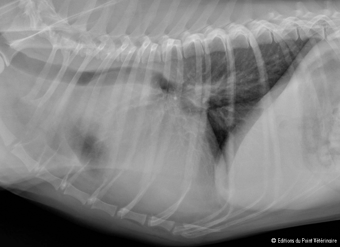 Syncope in a
Cavalier King Charles dog. C Fina, F
Stambouli. Point Veterinaire, 2011;42(316):9-10. Quote: The clinical
signs, diagnosis and surgical treatment of chemodectoma in a
Cavalier King Charles Spaniel dog in France are described.
Clinical presentation: A 7-year-old Cavalier King Charles male dog is
presented for consultation for the exploration of a heart murmur. The
owners report an episode of syncope 2 months before the consultation and
the appearance of a dry cough for 1 week. The clinical examination
confirms left apexal systolic systolic murmur (IV / VI) without any
other abnormality. X-rays of front and thorax profile are made (photos 1
and 2) .Quality of the image → The density, the contrast, the sharpness
and the fineness of the details are ...
Syncope in a
Cavalier King Charles dog. C Fina, F
Stambouli. Point Veterinaire, 2011;42(316):9-10. Quote: The clinical
signs, diagnosis and surgical treatment of chemodectoma in a
Cavalier King Charles Spaniel dog in France are described.
Clinical presentation: A 7-year-old Cavalier King Charles male dog is
presented for consultation for the exploration of a heart murmur. The
owners report an episode of syncope 2 months before the consultation and
the appearance of a dry cough for 1 week. The clinical examination
confirms left apexal systolic systolic murmur (IV / VI) without any
other abnormality. X-rays of front and thorax profile are made (photos 1
and 2) .Quality of the image → The density, the contrast, the sharpness
and the fineness of the details are ...
Case example: Collapse of a Cavalier King Charles Spaniel. Jacqui Gilmour. InPractice. May 2012;34(1):16-17. Quote: A 10-year-old neutered female Cavalier King Charles Spaniel (CKCS) was presented for further investigation of intermittent collapse. The dog had been medicated with 0·4-mg/kg benazepril every 24 hours since a murmur had been detected at four years of age. One month previously, the dog had presented with intermittent collapse and signs suggestive of left-sided congestive heart failure (CHF), and a diagnosis of chronic valvular heart disease (CVHD) and CHF was made. Furosemide (1·6-mg/kg every 12 hours) and spironolactone (2·4-mg/kg every 24 hours) had been added to the treatment regimen, which resulted in resolution of the cough and tachypnoea but not the collapsing episodes. At the time of investigation, the dog had suffered five episodes of collapse; on each occasion it woke up, stood up and then fell down into lateral recumbency with rigid limbs. During the episodes, the dog appeared unaware of its surroundings and it recovered spontaneously after less than 30 seconds. There was no change in behaviour before or after these events and there was no jaw movement or hypersalivation. The owners reported that the dog was mildly exercise intolerant, had a resting respiratory rate of 16 breaths per minute and had always suffered stertorous breathing when asleep. ... The description of the collapsing episodes is more characteristic for cardiac syncope; the dog suffered a transient loss of consciousness and postural tone, which lasted for less than one minute, followed by spontaneous recovery. ... Most commonly, cardiac syncope occurs during exercise or exertion. ... In this case, the episodes occurred on rising from sleep. A similar presentation is reported to occur in humans without structural heart disease. The pathogenesis in these patients is due to bradycardia and/or hypotension due to an inadequate function of the control mechanisms that normally maintain arterial blood pressure (ie, vasovagal syncope). In dogs, vasovagal syncope is typically reported to occur following situations such as extreme excitement, lead-pulling and coughing; to the authors' knowledge, there are no clinical reports of postural/orthostatic syncope. ... The rhythm present in the dog's ECG is a sinus arrhythmia with a wandering pacemaker, which is an unusual finding in cases of CHF. Ordinarily, the high sympathetic tone seen in CHF results in a sinus rhythm or sinus tachycardia. ... The dog was referred to the veterinary cardiorespiratory centre and a second-hand pacemaker (Sigma VVIR; Medtronic) was fitted, which led to complete resolution of the syncope. Conclusions: Syncope in CVHD typically occurs secondarily to PH, tachy-arrhythmias, cardiac tamponade caused by left atrial rupture, iatrogenic volume depletion and vasodilation, or secondary to a reduction in cardiac output following increased demand on the heart (eg, excitement). In this case, the syncope was caused by sinus arrest but the aetiology of the arrhythmia remains unclear. The dog may have been suffering from sick sinus syndrome or atrial myocarditis, or arrhythmias were precipitated by the dog's underlying CVHD resulting in increased left atrial size and possible myocardial hypoxia. Alternatively, the arrhythmias may have been exacerbated by high vagal tone, a consequence of the brachycephalic nature of this animal and/or concurrent respiratory disease. In addition, sleep apnoea has been reported in people and English bulldogs, and has been associated with arrhythmias.
Holter Monitoring of Small Breed Dogs with Advanced Myxomatous Mitral Valve Disease with and without a History of Syncope. C.E. Rasmussen, T. Falk, A. Domanjko Petric, M. Schaldemose, N.E. Zois, S.G. Moesgaard, B. Åblad, H.Y. Nilsen, I. Ljungvall, K. Hoglund, J. Haggstrom, H.D. Pedersen, J.M. Bland and L.H. Olsen. J.Vet.Int.Med. Jan. 2014. Quote: "Background: Syncope is a transient loss of consciousness occasionally occurring in dogs with advanced myxomatous mitral valve disease (MMVD). Objective: (1) To study ECG changes during syncopal episodes in dogs with advanced MMVD and (2) to compare the occurrence of arrhythmias and changes in heart rate variability (HRV) between dogs with advanced MMVD with and without a history of syncope. Animals: Forty-three privately owned dogs (<15 kg) with advanced MMVD: 21 with [including 6 cavalier King Charles spaniels] and 22 without a history of syncope [including 10 CKCSs]. Methods: Prospective study with dogs recruited for an evaluation including history, physical examination, echocardiography, and arrhythmia and HRV analysis performed on 24-hour Holter recordings. Results: A syncopal episode was observed during Holter monitoring in 4 dogs: 3 dogs had sinus rhythm and 1 dog had sinus arrest followed by escape rhythm. An arrhythmia variable representing sinus arrhythmia was significantly lower in dogs with a history of syncope than in those without (P = .008). Eight of 26 HRV variables were significantly different between dogs with and without a history of syncope. Conclusions and Clinical Importance: Compared with dogs without a history of syncope, dogs with advanced MMVD and a history of syncope did not have a higher occurrence of arrhythmias, but had less sinus arrhythmia, and had changes in HRV variables representing decreased overall HRV, decreased parasympathetic, and increased sympathetic modulation of heart rate."
Serum cardiac troponin I in canine syncope and seizures. E. Dutton, J. Dukes-McEwan, P.J. Cripps. J. Vet. Cardiol. February 2017;19(1):24-34. Quote: Objective: To determine if serum cardiac troponin I (cTnI) concentration distinguishes between cardiogenic syncope and collapsing dogs presenting with either generalized epileptic seizures (both with and without cardiac disease) or vasovagal syncope. Animals: Seventy-nine prospectively recruited dogs [including nine cavalier King Charles spaniels], grouped according to aetiology of collapse: generalized epileptic seizures (group E)[two cavaliers out of 30], cardiogenic syncope (group C)[one cavalier out of 20], dogs with both epileptic seizures and cardiac disease (group B)[four cavaliers out of 9], vasovagal syncope (group V) or unclassified (group U)[two cavaliers out of 13]. Methods: Most patients had ECG (n = 78), echocardiography (n = 78) and BP measurement (n = 74) performed. Dogs with a history of intoxications, trauma, evidence of metabolic disorders or renal insufficiency (based on serum creatinine concentrations >150 μmol/L and urine specific gravity <1.030) were excluded. Serum cTnI concentrations were measured and compared between groups using non-parametric statistical methods. Multivariable regression analysis investigated factors associated with cTnI. Receiver operator characteristic curve analysis examined whether cTnI could identify cardiogenic syncope. Results: Median cTnI concentrations were higher in group C than E (cTnI: 0.165 [0.02-27.41] vs. 0.03 [0.01-1.92] ng/mL; p<0.05). Regression analysis found that serum cTnI concentrations decreased with increasing time from collapse (p=0.015) and increased with increasing creatinine concentration (p=0.028). Serum cTnI diagnosed cardiogenic syncope with a sensitivity of 75% and specificity of 80%. Conclusions: Patients collapsing with a cardiac cause have significantly increased troponin concentrations compared with epileptic or vasovagal patients; however, the overlap in troponin concentrations reduces the discriminatory power of the test in an individual dog. Therefore, troponin assays should not be used as stand-alone tests, but in combination with other diagnostic investigations, such as ECG and echocardiography. Future large prospective studies investigating the possible prognostic value of cTnI in syncopal dogs are warranted.
Epileptic Seizures Versus Syncope: Pathophysiology and Clinical Approach. Marios Charalambous, Sergio A. Gomes, Stella Papageorgiou, Massimo Orioles. Vet. Evid. February 2017;2(1). Quote: Generalised epileptic seizures and syncope are two syndromes with similar clinical manifestation and their differentiation can be quite challenging. The aim of this review is to use an evidence-based approach in differentiating these two syndromes through the comprehension of the pathophysiological mechanisms involved and their clinical signs. Both syndromes affect regions of the forebrain and consciousness level, although, different mechanisms are involved. Syncope is a paroxysmal event secondary to a short-term decrease in cerebral perfusion, oxygenation or essential nutrients delivery. Generalised epileptic seizure activity is defined as the clinical manifestation of transient paroxysmal disturbances in brain function secondary to an imbalance between excitatory and inhibitory neurotransmitters. Clinical criteria, including precipitating events, clinical signs preceding, during and following the episodes and event duration, can be used to differentiate the two syndromes. Although these criteria might be useful for the practitioner, definite conclusions should be precluded due to the lack of original research articles and weak evidence on this specific field.
ACVIM consensus statement guidelines for the diagnosis, classification, treatment, and monitoring of pulmonary hypertension in dogs. Carol Reinero, Lance C. Visser, Heidi B. Kellihan, Isabelle Masseau, Elizabeth Rozanski, Cecile Clercx, Kurt Williams, Jonathan Abbott, Michele Borgarelli, Brian A. Scansen. J. Vet. Intern. Med. February 2020; doi: 10.1111/jvim.15725. Quote: Pulmonary hypertension (PH), defined by increased pressure within the pulmonary vasculature, is a hemodynamic and pathophysiologic state present in a wide variety of cardiovascular, respiratory, and systemic diseases. The purpose of this consensus statement is to provide a multidisciplinary approach to guidelines for the diagnosis, classification, treatment, and monitoring of PH in dogs. Comprehensive evaluation including consideration of signalment, clinical signs, echocardiographic parameters, and results of other diagnostic tests supports the diagnosis of PH and allows identification of associated underlying conditions. ... Exertional syncope, labored respiration or overt respiratory distress, and exercise intolerance are noteworthy clinical signs frequently directly attributable to PH. ... In the veterinary medical literature, the degree or severity of PH has been classified as mild, moderate, or severe. These categories are based on the PG derived from TRV (also called the tricuspid regurgitation PG) or estimated systolic PAP. Cutoffs used for these categories (mild, moderate, severe) are arbitrary, and the categories are potentially misleading or flawed. For example, based on the conventional definition of PH (based solely on estimated systolic PAP), a dog with severe PH can be free of clinical signs, whereas a dog with moderate PH could be in rightsided heart failure. Therefore, the panel does not advocate their use. Instead, severity of PH should be based on clinical findings (Table 1) and outcome data from large (prospective) longitudinal studies, which unfortunately are unavailable. ... TABLE 1: Clinical findings suggestive of pulmonary hypertension (PH) in dogs -- Findings strongly suggestive of PH -- Syncope (especially with exertion or activity) without another identifiable cause. ... The panel recognizes the probabilistic approach to diagnosis of PH presents challenges, particularly regarding standardized enrollment in future clinical studies of dogs with suspected PH. Therefore, we propose that the clinical definition of PH should include dogs with intermediate or high probability of PH, and specifically, a tricuspid regurgitation PG cutoff of >46 mm Hg (TRV >3.4 m/s). This roughly equates to what has been conventionally referred to as at least moderate PH. Until there are echocardiographic variables that are repeatable and have acceptable diagnostic accuracy relative to RHC, this cutoff value could be used with an understanding of its limitations. ... Dogs with PH can be classified into the following 6 groups: group 1, pulmonary arterial hypertension; group 2, left heart disease; group 3, respiratory disease/hypoxia; group 4, pulmonary emboli/pulmonary thrombi/pulmonary thromboemboli; group 5, parasitic disease (Dirofilaria and Angiostrongylus); and group 6, disorders that are multifactorial or with unclear mechanisms. The approach to treatment of PH focuses on strategies to decrease the risk of progression, complications, or both, recommendations to target underlying diseases or factors contributing to PH, and PH-specific treatments. ... Treatment strategies for targeting the underlying disease in group 2 patients are centered around identifying and, if possible, reversing the cause of LHD [left-sided heart disease], decreasing postcapillary PH (ie, lowering LA pressure) and, if present, treating heart failure. ... Because dogs with PH secondary to LHD, by definition, have postcapillary PH (with or without pre-PH), the use of phosphodiesterase 5 inhibitors (PDE5i) [e.g., sildenafil (Viagra)] is not recommended as first line treatment. ... A PDE5i should be administered only to dogs free of acute or decompensated LHF (cardiogenic pulmonary edema). Heart failure medications (HFM) and a PDE5i are recommended for dogs with clinical (eg, jugular venous distension, fluid wave on abdominal palpation, or auscultable pleural fluid line) and ultrasonographic (abdominal or pleural effusion without another cause along with RA enlargement, caudal vena caval distension, hepatic venous distension or hepatomegaly) evidence of right-sided heart failure. Addition of a PDE5i may be considered in dogs with exertional syncope without another identifiable cause that have failed to respond to other treatments for preclinical LHD (eg, pimobendan). ... A PDE5i may be considered for dogs with compensated LHF and a high probability of PH (ie, dogs previously diagnosed with LHF and on HFM but that do not currently have pulmonary edema) that develop exertional syncope without another identifiable cause. A thorough diagnostic evaluation is advised to rule out alternative causes of syncope (eg, ECG, ambulatory ECG monitoring, or both to exclude bradyarrhythmias or tachyarrhythmias as potential causes for syncope). ... Dogs with PH should be monitored for improvement, static condition, or progression, and any identified underlying disorder should be addressed and monitored simultaneously.
Atypical vasovagal syncope in cavalier King Charles spaniel ocurring at rest. T. Lahuerta-Smith, M.-L. Theron. Revue Veterinaire Clinique. April 2021; doi: 10.1016/j.anicom.2021.02.001. Quote: A 10-year-old male entire Cavalier King Charles Spaniel was referred for investigation of frequent episodes of transient loss of consciousness during periods of rest as well as severe stertor and inspiratory dyspnoea. Echocardiography revealed degenerative mitral valve disease in stage B1 ACVIM (Keene et al., 2019) without any evidence of pulmonary hypertension. Respiratory tract endoscopic examination revealed a significantly enlarged soft palate, laryngeal saccule eversion and severe laryngeal collapse. During syncopal episodes, Holter examination revealed progressive bradycardia with long periods of sinus and ventricular arrest, followed by paroxysmal runs of atrial fibrillation, suggestive of reflex syncope. This case describes a dog with reflex syncope occurring in an atypical situation of rest with spontaneous improvement of syncope frequency until its complete resolution 6 months after presentation. Before considering pacemaker implantation to improve clinical signs, medical therapy was initiated with theophylline at 5 mg/kg twice daily orally (Dilatrane, SERP), in an effort to increase cardiac sympathetic tone and limit vagal stimulation on the heart. Treatment was stopped two weeks later as there was no obvious clinical benefit following medical therapy, with various syncopal episodes occurring daily. One month later, the animal had a significant reduction in syncopal frequency (around 5 per day), as well as a change in the nature of the episodes: these were shorter in duration, the dog did not lose postural tone but rather fell on its hindlimbs, and there was no loss of consciousness, which was suggestive of near-syncope. Six months later after initial presentation and surgery, the dog no longer has syncopal episodes of any kind. A control Holter was obtained at this time, which showed a normal sinus rhythm throughout the day with a complete absence of cardioinhibitory reflexes.
CONNECT WITH US